Proportion of Hosts Carrying Batrachochytrium
dendrobatidis, Causal Agent of Amphibian Chytridiomycosis,
in Oswego County, NY in 2012
Sofia T. Windstam and Jennifer C. Olori
Northeastern Naturalist, Volume 21, Issue 1 (2014): NENHC-25—NENHC-34
Full-text pdf (Accessible only to subscribers. To subscribe click here.)
Access Journal Content
Open access browsing of table of contents and abstract pages. Full text pdfs available for download for subscribers.
Current Issue: Vol. 30 (3)
Check out NENA's latest Monograph:
Monograph 22


Northeastern Naturalist Vol. 21, No. 1
S.T. Windstam and J.C. Olori
2014
NENHC-25
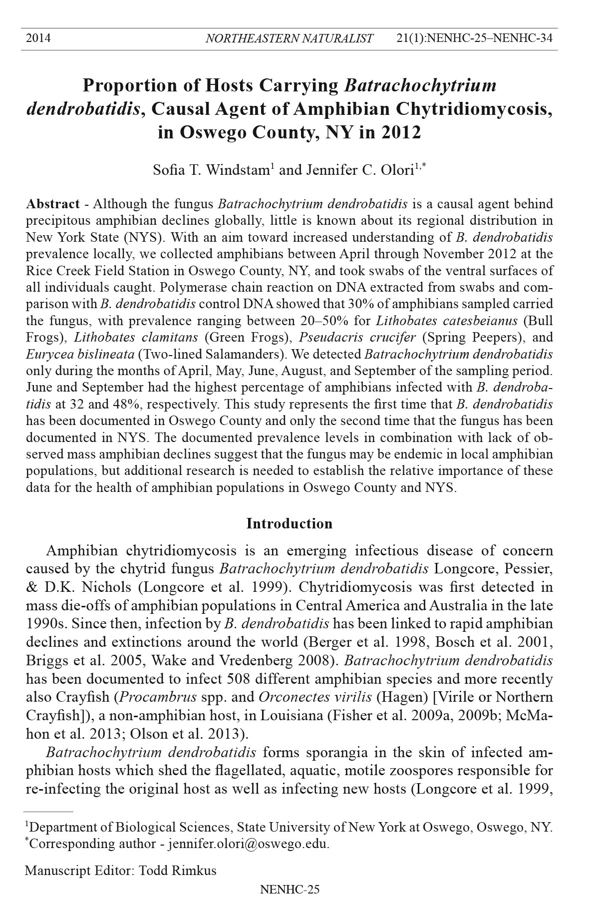
2014 NORTHEASTERN NATURALIST 21(1):NENHC-25–NENHC-34
Proportion of Hosts Carrying Batrachochytrium
dendrobatidis, Causal Agent of Amphibian Chytridiomycosis,
in Oswego County, NY in 2012
Sofia T. Windstam1 and Jennifer C. Olori1,*
Abstract - Although the fungus Batrachochytrium dendrobatidis is a causal agent behind
precipitous amphibian declines globally, little is known about its regional distribution in
New York State (NYS). With an aim toward increased understanding of B. dendrobatidis
prevalence locally, we collected amphibians between April through November 2012 at the
Rice Creek Field Station in Oswego County, NY, and took swabs of the ventral surfaces of
all individuals caught. Polymerase chain reaction on DNA extracted from swabs and comparison
with B. dendrobatidis control DNA showed that 30% of amphibians sampled carried
the fungus, with prevalence ranging between 20–50% for Lithobates catesbeianus (Bull
Frogs), Lithobates clamitans (Green Frogs), Pseudacris crucifer (Spring Peepers), and
Eurycea bislineata (Two-lined Salamanders). We detected Batrachochytrium dendrobatidis
only during the months of April, May, June, August, and September of the sampling period.
June and September had the highest percentage of amphibians infected with B. dendrobatidis
at 32 and 48%, respectively. This study represents the first time that B. dendrobatidis
has been documented in Oswego County and only the second time that the fungus has been
documented in NYS. The documented prevalence levels in combination with lack of observed
mass amphibian declines suggest that the fungus may be endemic in local amphibian
populations, but additional research is needed to establish the relative importance of these
data for the health of amphibian populations in Oswego County a nd NYS.
Introduction
Amphibian chytridiomycosis is an emerging infectious disease of concern
caused by the chytrid fungus Batrachochytrium dendrobatidis Longcore, Pessier,
& D.K. Nichols (Longcore et al. 1999). Chytridiomycosis was first detected in
mass die-offs of amphibian populations in Central America and Australia in the late
1990s. Since then, infection by B. dendrobatidis has been linked to rapid amphibian
declines and extinctions around the world (Berger et al. 1998, Bosch et al. 2001,
Briggs et al. 2005, Wake and Vredenberg 2008). Batrachochytrium dendrobatidis
has been documented to infect 508 different amphibian species and more recently
also Crayfish (Procambrus spp. and Orconectes virilis (Hagen) [Virile or Northern
Crayfish]), a non-amphibian host, in Louisiana (Fisher et al. 2009a, 2009b; McMahon
et al. 2013; Olson et al. 2013).
Batrachochytrium dendrobatidis forms sporangia in the skin of infected amphibian
hosts which shed the flagellated, aquatic, motile zoospores responsible for
re-infecting the original host as well as infecting new hosts (Longcore et al. 1999,
1Department of Biological Sciences, State University of New York at Oswego, Oswego, NY.
*Corresponding author - jennifer.olori@oswego.edu.
Manuscript Editor: Todd Rimkus
Northeastern Naturalist
NENHC-26
S.T. Windstam and J.C. Olori
2014 Vol. 21, No. 1
Rosenblum et al. 2010, Wake and Vredenberg 2008). Zoospore locomotion relies
on the presence of free-water films. The fungal infection is restricted to keratin
present in the epidermis of adult amphibians and larval salamanders and also in the
mouthparts of tadpoles. Although in tadpoles the specific effect of B. dendrobatidis
infection varies by species (Blaustein et al. 2005), tadpoles often do not die as a direct
result of infection, and the infection is lost at metamorphosis. However, newly
metamorphosed individuals can become re-infected and then die (Carey et al. 2003).
Adults of different host species also vary in their susceptibility to B. dendrobatidis;
Lithobates catesbeianus (Shaw) (Bullfrog) and Ambystoma tigrinum Green (Tiger
Salamander), both native to NYS, sometimes are considered to be reservoirs or
vectors because they may carry fungal infections without suffering from chytridiomycosis
(Carey et al. 2003, Gahl et al. 2011, Searle et al. 2011; but see Gervasi
et al. 2013). However, in many species, the mortality rate following infection is
55–99% of the population (Carey et al. 2003). The pathogen causes a characteristic
thickening of the stratum corneum (upper-most layer of epidermis) that disrupts the
ability of infected animals to regulate ion transport across the skin, ultimately causing
decreased blood-plasma potassium and sodium levels that leads to cardiac arrest
(Voyles et al. 2009). However, there are also indications that B. dendrobatidis may
release some sort of a chemical that is partially responsible for the observed host
pathology (Carey et al. 2003, McMahon et al. 2013).
Amphibian chytridiomycosis is unusual not only because fungi of the phylum
Chytridiomycota rarely are severe animal pathogens, but because B. dendrobatidis
is the first chytrid fungus known to infect vertebrates (Longcore et al. 1999).
Furthermore, it is currently not known if chytridiomycosis in amphibians has become
widespread because of a recent change in virulence of B. dendrobatidis or
susceptibility of its amphibian hosts (perhaps caused by environmental changes
or anthropogenic stressors). Another possibility is that the fungus was recently introduced
and has since spread to multiple new regions (Carey et al. 2003, Kilpatrick
et al. 2009, Lips et al. 2008).
Despite the presence of B. dendrobatidis in Quebec, Canada (Ouelett et al.
2005), and the Northeast US (Longcore et al. 2007), specimens from NYS were
not reported to test positive for the presence of B. dendrobatidis until 2012 (Becker
et al. 2012), and amphibian declines connected to chytridiomycosis have not been
described in the Northeast. Current hypotheses suggest that either declines are ongoing
but have not been detected because of a lack of long-term population data
in the Northeast, or that the absence of declines are real and may result from lessvirulent
local strains of B. dendrobatidis or a natural resistance to the pathogen in
at least some local species (Gahl et al. 2011). Moreover, results from recent studies
in the Southeast suggested strong seasonality in the prevalence of B. dendrobatidis
infections, with peak prevalence (45% of individuals) occurring in the spring,
whereas levels are minimal in the fall (2%) (Pullen et al. 2010 ).
The apparent lack of widespread, pathogenic outbreaks of chytridiomycosis and
mass-die offs of amphibians in the Northeast also could result from a combination
of those factors proposed above, such that a local strain of B. dendrobatidis
Northeastern Naturalist Vol. 21, No. 1
S.T. Windstam and J.C. Olori
2014
NENHC-27
may be controlled by climactic and seasonal variables specific to the Northeast in
conjunction with the resistance of at least some native species (Gahl et al. 2011).
However, because local amphibian population trends are not actively monitored in
many areas, and local species have not been tested regularly for the presence and
prevalence of B. dendrobatidis, it is unknown which or how many strains could be
present in the area, and little information exists on the annual pathogenicity and
transmission patterns of B. dendrobatidis in the Northeast.
Our goal was to investigate the prevalence of B. dendrobatidis infection in local
populations of amphibians in order to address the following questions: 1) Is
B. dendrobatidis present in local populations? 2) Do patterns of prevalence in the
Northeast reflect similar seasonal patterns detected in the Sout heast? The first step
in this long-term project was to address whether or not B. dendrobatidis presently
is infecting local amphibians.
Materials and Methods
Field-site description and sampling of amphibians
We sampled a total of 82 amphibians representing five different species (6
Bull Frogs, 49 Lithobates clamitans Latreille [Green Frogs], 1 Lithobates pipiens
(Schreber) [Northern Leopard Frog], 5 Pseudacris crucifer (Wied-Neuwied)
[Spring Peepers], and 16 Eurycea bislineata (Green) [Two-lined Salamanders])
over 11 visits to the SUNY Oswego Rice Creek Field Station (RCFS) between the
months of April through November 2012 (Fig. 1). The majority of RCFS is abandoned
agricultural land that is being allowed to undergo natural succession, with
Figure 1. Number of individuals of each species of amphibian sa mpled by month.
Northeastern Naturalist
NENHC-28
S.T. Windstam and J.C. Olori
2014 Vol. 21, No. 1
different areas at various stages of succession represented (Weeks and Cox 1988).
The site contains multiple wetlands (e.g., large pond, creek, marshes, and vernal
pools), meadows, old woodlots, and hardwood forest. All sampled individuals were
adults or sub-adults, and the vast majority of animals sampled were collected from
or adjacent to the pond, creek, or vernal pools. We captured animals either using
nets or by hand with sterile nitrile gloves. We then identified individuals to species
and swabbed (Medical Wire and Equipment Co., UK) each one five times along
each of the ventral surfaces of the hands, legs, feet, abdomen, thighs, and pelvic
patch. Swab tips were broken off into sterile 1.5-ml microcentrifuge tubes containing
one ml of 70% ethanol, which were then stored at -4 ºC until processed. We
also collected standard data such as snout–vent length, weight, sex, and condition
(external appearance and overall health), along with environmental information
(temperature, humidity, weather, etc.) for each swabbed individual and entered the
information into a database for long-term population monitoring. During sampling,
we placed each amphibian into a new plastic bag to prevent cross-contamination
of equipment, and all gloves were changed and all equipment was sterilized using
either 10% bleach solution or 70% ethanol after each individual was processed.
DNA extraction
We transferred swabs to sterile 2-ml polypropylene tubes (Biospec, OK) containing
30–40 mg of sterile 0.5-mm zirconium/silica beads (Biospec). We added
fifty μl of PrepMan Ultra DNA extraction solution (Applied Biosystems, CA)
to each tube and homogenized the samples for 50 s using a Mini-Beadbeater-1
(Biospec). We then centrifuged the samples for 30 s at 13,000 × g, after which
we repeated homogenization and spinning of the samples. Thereafter, samples
were boiled for 10 min, cooled for 2 min on ice, and centrifuged for 3 min at
13,000 × g. We collected 20 μl of the resulting supernatant to be stored long term
at -80 ºC (Hyatt et al. 2007).
PCR amplification and gel electrophoresis
Although quantitative PCR (qPCR) has been the standard when diagnosing B.
dendrobatidis infections of amphibians, a recent publication demonstrated that
conventional end-point PCR is equally sensitive in detecting B. dendrobatidis and
therefore is appropriate for analyses in which prevalence data are collected (Boyle
et al. 2004, Garland et al. 2011). We diluted all DNA extracts tenfold in molecular
grade water prior to subjecting them to PCR. PCR reactions contained 0.8 u Fast-
Start Taq polymerase (Roche Applied Science, IN), 0.25 mM each of dATP, dCTP,
dGTP, and dTTP (Invitrogen, Life Technologies, NY), 0.9 μM forward primer
ITS1-3 Chytr (5’-CCTTGATATAATACAGTGTGCCATATGTC-3’, Invitrogen),
0.9 μM reverse primer 5.8S Chytr (5’-AGCCAAGAGATCCGTTGTCAAA-3’,
Invitrogen), 3 mM MgCl2, 4 μl diluted DNA template, and sterile molecular-grade
water to achieve a final reaction volume of 20 μl (Boyle et al. 2004, Garland et al.
2011). We performed PCR using the following temperature protocol: initial denaturation
at 95 ºC for 4 min followed by 50 cycles of denaturing at 95 ºC for 30 s,
annealing at 55 ºC for 30 s, and extension at 72 ºC for 45 s. We carried out all PCR
Northeastern Naturalist Vol. 21, No. 1
S.T. Windstam and J.C. Olori
2014
NENHC-29
reactions in individual 0.2-ml PCR tubes, and each round of PCR included a negative
and a positive control using sterile molecular-grade water and DNA isolated
from B. dendrobatidis cultures, respectively. We mixed 10-μl aliquots of PCR products
with loading dye and analyzed them using 2% agarose gels in 1X TAE buffer.
The positive control DNA from B. dendrobatidis generated an anticipated band
size of 146 bp, and we deemed samples to be positive for the presence of B. dendrobatidis
if: 1) controls yielded the expected results (no amplification in negative
control and amplification in positive control) and 2) the sample produced a band of
the anticipated size. Band sizes were ascertained by comparing migrations of DNA
fragments in a 100-bp DNA ladder (Fisher BioReagents, PA) as well as comparing
to the relative band location of the positive control.
Results and Discussion
Batrachochytrium dendrobatidis was detected in 30% of the sampled amphibians
(data not shown). The fungus was detected on Green Frogs, Bull Frogs, Spring
Peepers, and Two-lined Salamanders, with a prevalence of 33, 50, 20, and 20%,
respectively (Fig. 2). The prevalence levels for Bull Frogs and Spring Peepers in
particular have to be viewed as tentative due to the low number of animals sampled
(6 Bull Frogs and 5 Spring Peepers). Likewise, the absence of B. dendobatidis on
Northern Leopard Frogs has to be viewed with similarly guarded skepticism due to
only one sampled individual. Survey data of B. dendrobatidis prevalence levels for
Two-lined Salamanders at other Northeast sites is lacking, and our study presents
a preliminary glance at prevalence levels of the chytrid fungus on this amphibian.
A recently published survey in the Adirondacks, NY, found B. dendrobatidis to be
Figure 2. Overall B. dendrobatidis prevalence rates on all amphibian species found to test
positive for the fungus.
Northeastern Naturalist
NENHC-30
S.T. Windstam and J.C. Olori
2014 Vol. 21, No. 1
present on approximately 25% of Green Frogs sampled in the latter part of June at
two pond sites (Becker et al. 2012). Thirty-three percent of Green Frogs at RCFS
tested positive for Bd, demonstrating that 25–33% of Green Frogs carry B. dendrobatidis
at two geographically distinct locations in NYS. How indicative these
B. dendrobatidis prevalence levels are among Green Frogs in NYS remains to be
established, as this study along with Becker et al. (2012) are the only studies to date
that have examined B. dendrobatidis prevalence.
The significance of B. dendrobatidis prevalence levels is not yet clear because
of the lack of published reports of amphibian population declines connected to
chytridiomycosis in the Northeast. It is possible that population declines are ongoing,
declines have already taken place, or the lack of decline is actually real.
Lack of current observable population declines could result from one or a combination
of factors, including the introductory wave of infection having already swept
through the amphibian population locally, the presence of less virulent local isolates
of B. dendrobatidis in NYS, some level of protection afforded by local environmental
or climatic conditions, or a higher natural resistance against the pathogen in
our local populations (Gahl et al. 2011). Using Green Frogs as an example, Gahl et
al. (2011) demonstrated that these particular amphibians are susceptible to chytrid
infection and die at appreciable rates once infected, but that there exists an isolatespecific
effect. Green Frogs were more susceptible to a novel isolate originating
from Panama, and not at all sensitive to a strain isolated from the Northeast (Gahl
et al. 2011). Consequently, in order to tease apart whether observed B. dendrobatidis
prevalence levels may be linked to any past or future declines it will be
critical to explore which isolates of B. dendrobatidis are present locally in order to
ascertain potential effects of the fungus on local amphibian populations. Lack of
disease may simply be a matter of local isolates being less virulent, but until this
has been established experimentally, it remains mere speculation. Obtaining local
B. dendrobatidis isolates would also allow for further controlled studies on potential
modulating effects of climatic conditions on infection outcomes. Furthermore,
resistance in some amphibians against B. dendrobatidis may be conferred by resident
skin-microbe populations (Harris et al. 2006, 2009a, 2009b), but it is unclear
whether or not such resistance mechanisms are in place in our local populations,
and this possibility remains to be explored.
Until recently, NYS had no published records of amphibians testing positive for
the presence of B. dendrobatidis (Becker et al. 2012). Our study represents the first
time that B. dendrobatidis has been documented in amphibians of Oswego County.
In Virginia, a strong seasonality has been described for B. dendrobatidis prevalence
at six different sites in both rural and urban areas, where rates of amphibians carrying
B. dendrobatidis peaked in spring between March–June and declined to 2%
in the fall (Pullen et al. 2010). In NYS, it is unclear whether such seasonality is
expected to take place, but our data suggest that the prevalence of B. dendrobatidis
fluctuates, which may be a random pattern or the result of seasonality. Potentially
seasonal effects could simply be a function of physiology because B. dendrobatidis
displays optimal growth at 17–25 °C, with temperatures below 5 °C and at or above
Northeastern Naturalist Vol. 21, No. 1
S.T. Windstam and J.C. Olori
2014
NENHC-31
28 °C significantly restricting growth (Piotrowski et al. 2004). We detected B. dendrobatidis
on amphibians only during April, May, June, August, and September of
the sampling period, during which June and September had the highest proportions
of amphibians infected with B. dendrobatidis at 32 and 48%, respectively (Fig. 3).
Data from the Adirondacks in NYS provides plausible support for the effect of
temperature on infection because a site with increased canopy cover experiencing
lower temperatures had higher B. dendrobatidis infection loads on Green Frogs
than individuals from a site with less cover and increased temperature (Becker et
al. 2012). Considering the effect temperature has on B. dendrobatidis physiology
it stands to reason that NYS, with a climate that experiences significant seasonal
variation in temperature, would exhibit some form of seasonal variation (Becker et
al. 2012, Piotrowski et al. 2004, Pullen et al. 2010). However, additional data from
multiple years and sites locally are necessary prior to inferring with certainty the
nature of these fluctuations.
This study is the first documentation of B. dendrobatidis in Oswego County in
NYS and represents a first step in addressing the effect this pathogen may have on
our local amphibian populations. In order to interpret the significance of our observations,
additional research on many aspects of infection is required, including
continued monitoring and surveying of amphibian populations for B. dendrobatidis
across different seasons. For future work, isolation and description of the fungal
isolates present locally is necessary in order to determine pathogen population
variation and structure, which are vital for explaining variation in pathogenicity
and virulence should this be an underlying factor that explains lack of observed
population declines (Fisher et al. 2009a, Gahl et al. 2011). The genetic population
structure of B. dendrobatidis may also provide information on whether or not the
Figure 3. Prevalence rate of B. dendrobatidis on all sampled amphibians per month sampled.
Northeastern Naturalist
NENHC-32
S.T. Windstam and J.C. Olori
2014 Vol. 21, No. 1
fungus was recently introduced regionally as opposed to displaying some endemism
(Morgan et al. 2007).
Acknowledgments
Positive control DNA extracted from B. dendrobatidis cultures was very generously
donated by Dr. Kelly Zamudio at the Department of Ecology and Evolutionary Biology at
Cornell University. This research was supported by a grant from the Rice Creek Associates
to J.C. Olori and S.T. Windstam. We are indebted to the numerous undergraduate students,
staff, and faculty members who have participated in the field sampling of amphibians and
molecular diagnostics.
Literature Cited
Becker, C.G., D. Rodriguez, A.V. Longo, A.L. Talaba, and K.R. Zamudio. 2012. Disease
risk in temperate amphibian populations is higher at closed-canopy sites. PLoS One
7:e48205.
Berger, L., R. Speare, P. Daszak, D.E. Green, A.A. Cunningham, C.L. Goggin, R. Slocombe,
M.A. Ragan, A.D. Hyatt, K.R. McDonald, H.B. Hines, K. R. Lips, G. Mantelli,
and H. Parkes. 1998. Chytridiomycosis causes amphibian mortality associated with
population declines in the rain forests of Australia and Central America. Proceedings of
the National Academy of Sciences, USA 95:9031–9036.
Blaustein, A.R., J.M. Romansic, E.A. Scheessele, B.A. Han, A.P. Pessier, and J.E. Longcore.
2005. Interspecific variation in susceptibility of frog tadpoles to the pathogenic
fungus Batrachochytrium dendrobatidis. Conservation Biology 19:1460–1468.
Bosch, J., I. Martinez-Solano, and M. Garcia-Paris. 2001. Evidence of a chytrid fungus
infection involved in the decline of the Common Midwife Toad (Alytes obstetricans) in
protected areas in central Spain. Biological Conservation 97:33 1–337.
Boyle, D.G., D.B. Boyle, V. Olsen, J.A.T. Morgan, and A.D. Hyatt. 2004. Rapid quantitative
detection of chytridiomycosis (Batrachochytrium dendrobatidis) in amphibian samples
using real-time Taqman PCR assay. Diseases of Aquatic Organisms 60:141–148.
Briggs, C.J., V.T. Vredenburg, R. Knapp, and L.J. Rachowicz. 2005. Investigating the
population-level effects of chytridiomycosis: An emerging infectious disease of amphibians.
Ecology 86:3149–3159.
Carey, C., A.P. Pessier, and A.D. Peace. 2003. Pathogens, infectious disease, and immune
defenses. Pp. 127–136, In R.D. Semlitsch (Ed.). Amphibian Conservation. Smithsonian
Books, Washington, DC.
Fisher, M.C., J. Bosch, Z. Yin, D.A. Stead, J. Walker, L. Selway, A.J.P. Brown, L.A. Walker,
N.A.R. Gow, J.E. Staijch, and T.W.J. Garner. 2009a. Proteomic and phenotypic profiling
of the amphibian pathogen Batrachochytrium dendrobatidis shows that genotype is
linked to virulence. Molecular Ecology 18:415–429.
Fisher, M.C., T.W.J. Garner, and S.F. Walker. 2009b. Global emergence of Batrachochytrium
dendrobatidis and amphibian chytridiomycosis in space, time, and host. Annual
Review of Microbiology 63:291–310.
Gahl, M.K., J.E. Longcore, and J.E. Houlahan. 2011. Varying responses of northeastern
North American amphibians to the chytrid pathogen Batrachochytrium dendrobatidis.
Conservation Biology 26:135–141.
Garland, S., J. Wood, and L.F. Skerratt. 2011. Comparison of sensitivity between real-time
detection of a Taqman assay for Batrachochytrium dendrobatidis and conventional detection.
Diseases of Aquatic Organisms 94:101–105.
Northeastern Naturalist Vol. 21, No. 1
S.T. Windstam and J.C. Olori
2014
NENHC-33
Gervasi, S.S., J. Urbina, J. Hua, T. Chestnut, R.A. Relyea, and A.R. Blaustein. 2013. Experimental
evidence for American Bullfrog (Lithobates catesbeianus) susceptibility to
chytrid fungus (Batrachochytrium dendrobatidis). EcoHealth 10:166–171.
Harris, R.N., T.Y. James, A. Lauer, M.A. Simon, and A. Patel. 2006. Amphibian pathogen
Batrachochytrium dendrobatidis is inhibited by the cutaneous bacteria of amphibian
species. EcoHealth 3:53–56.
Harris, R.N., R.M. Brucker, J.B. Walke, M.H. Becker, C.R. Schwantes, D.C. Flaherty, B.A.
Lam, D.C. Woodhams, C.J. Briggs, V.T. Vredenburg, and K.P.C Minbiole. 2009a. Skin
microbes on frogs prevent morbidity and mortality caused by a lethal skin fungus. ISME
Journal 3:818–824.
Harris, R.N., A. Lauer, M.A. Simon, J.L. Banning, and R.A. Alford. 2009b. Addition of
antifungal skin bacteria to salamanders ameliorates the effects of chytridiomycosis.
Diseases of Aquatic Organisms 83:11–16.
Hyatt, A.D., D.G. Boyle, V. Olsen, D.B. Boyle, L. Berger, D. Obendorf, A. Dalton, K.
Kriger, M. Hero, H. Hines, R. Phillott, R. Campbell, G. Marantelli, F. Gleason, and A.
Colling. 2007. Diagnostic assays and sampling protocols for the detection of Batrachochytrium
dendrobatidis. Diseases of Aquatic Organisms 73:175–192.
Kilpatrick, A.M., C.J. Briggs, and P. Daszak. 2009. The ecology and impact of chytridiomycosis:
an emerging disease of amphibians. Trends in Ecology and Evolution 25:109–118.
Lips K.R., J. Diffendorfer, J.R. Mendelson III, and M.W. Sears. 2008. Riding the wave:
Reconciling the roles of disease and climate change in amphibian declines. PLoS Biology
6:e72.
Longcore, J.E., A.P. Pessier, and D.K. Nichols. 1999. Batrachochytrium dendrobatidis gen.
et sp. nov., a chytrid pathogenic to amphibians. Mycologia 91:219–227.
Longcore, J.R., J.E. Longcore, A.P. Pessier, and W.A. Halteman. 2007. Chytridiomycosis
widespread in anurans of northeastern United States. Journal of Wildlife Management
71:435–444.
McMahon, T.A., L.A. Brannelly, M.W.H. Chatfield, P.T.J. Johnson, M.B. Joseph, V.J.
McKenzie, C.L. Richards-Zawacki, M.D. Venesky, and J.R. Rohr. 2013. Chytrid fungus
Batrachochytrium dendrobatidis has nonamphibian hosts and releases chemicals that
cause pathology in the absence of infection. Proceedings of the National Academy of
Sciences, USA 110:210–215.
Morgan, J.A.T., V.T. Vredenburg, L.J. Rachowicz, R.A. Knapp, M.J. Stice, T. Tunstall, R.E.
Bingham, J.M. Parker, J.E. Longcore, C. Moritz, C.J. Briggs, and J.W. Taylor. 2007.
Population genetics of the frog-killing fungus Batrachochytrium dendrobatidis. Proceedings
of the National Academy of Sciences, USA 104:13,845–13,850.
Olson, D.H., D.M. Aanensen, K.L. Ronnenberg, C.I. Powell, S.F. Walker, J. Bielby, T.W.J.
Garner, G. Weaver, M.C. Fisher, and the Bd Mapping Group. 2013. Mapping the global
emergence of Batrachochytrium dendrobatidis, the amphibian chytrid fungus. PLoS
ONE 8:e56802.
Ouelett, M., I. Mikaelian, B.D. Pauli, J. Rodrigue, and D.M. Green. 2005. Historical evidence
of widespread chytrid infection in North American amphibian populations. Conservation
Biology 19:1431–1440.
Piotrowski, J.S., S.L. Annis, and J.E. Longcore. 2004. Physiology of Batrachochytrium
dendrobatidis, a chytrid pathogen of amphibians. Mycologia 96:9–15.
Pullen K.D., A.M. Best, and J.L. Ware. 2010. Amphibian pathogen Batrachochytrium dendrobatidis
prevalence is correlated with season and not urbanization in central Virginia.
Diseases of Aquatic Organisms 91:9–16.
Northeastern Naturalist
NENHC-34
S.T. Windstam and J.C. Olori
2014 Vol. 21, No. 1
Rosenblum, E.B., J. Voyles, T.J. Poorten, and J.E. Stajich. 2010. The deadly chytrid fungus:
A story of an emerging pathogen. PLoS Pathogens 6:e1000550.
Searle, C.L., S.S. Gervasi, J. Hua, J.I. Hammond, R.A. Relyea, D.H. Olson, and A.R.
Blaustein. 2011. Differential host susceptibility to Batrachochytrium dendrobatidis, an
emerging amphibian pathogen. Conservation Biology 25:965–974.
Voyles, J., S. Young, L. Berger, C. Campbell, W.F. Voyles, A. Dinudom, D. Cook, R. Webb,
R.A. Alford, L.F. Skerratt, and R. Speare. 2009. Pathogenesis of chytridiomycosis, a
cause of catastrophic amphibian declines. Science 326:582–585.
Wake, D.B., and V.T. Vredenberg. 2008. Are we in the midst of the sixth mass extinction? A
view from the world of amphibians. Proceedings of the National Academy of Sciences,
USA 105:11,466–11,473.
Weeks, J.A., and D.D. Cox. 1988. Guidelines for environmental management at Rice Creek
Field Station. Rice Creek Field Station Bulletin No. 6. 42 pp. + appendices.