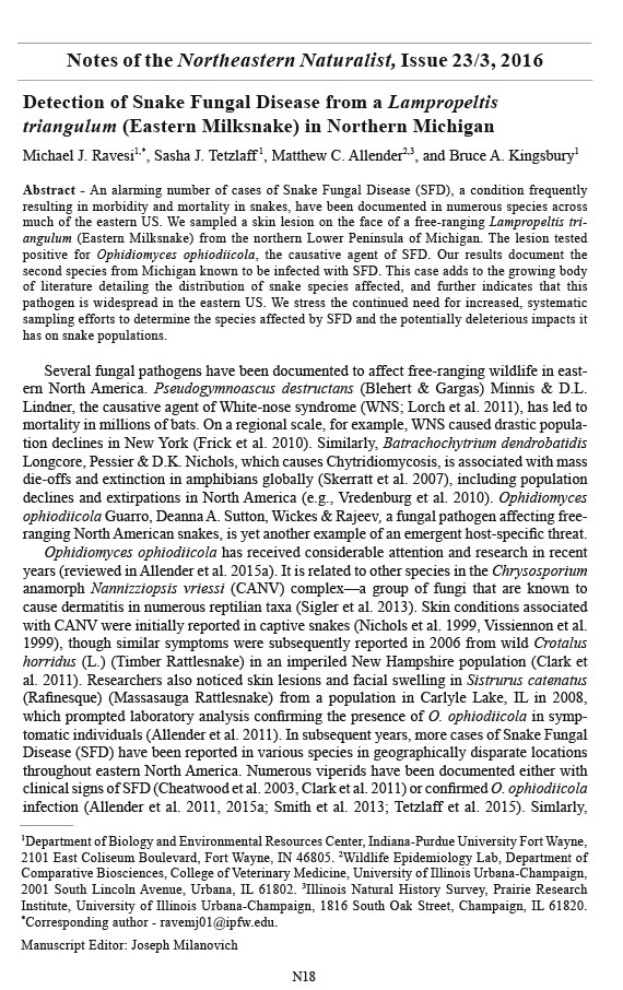
Detection of Snake Fungal Disease from a Lampropeltis
triangulum (Eastern Milksnake) in Northern Michigan
Michael J. Ravesi, Sasha J. Tetzlaff, Matthew C. Allender, and Bruce A. Kingsbury
Northeastern Naturalist, Volume 23, Issue 3 (2016): N18–N21
Full-text pdf (Accessible only to subscribers. To subscribe click here.)
Access Journal Content
Open access browsing of table of contents and abstract pages. Full text pdfs available for download for subscribers.
Current Issue: Vol. 30 (3)
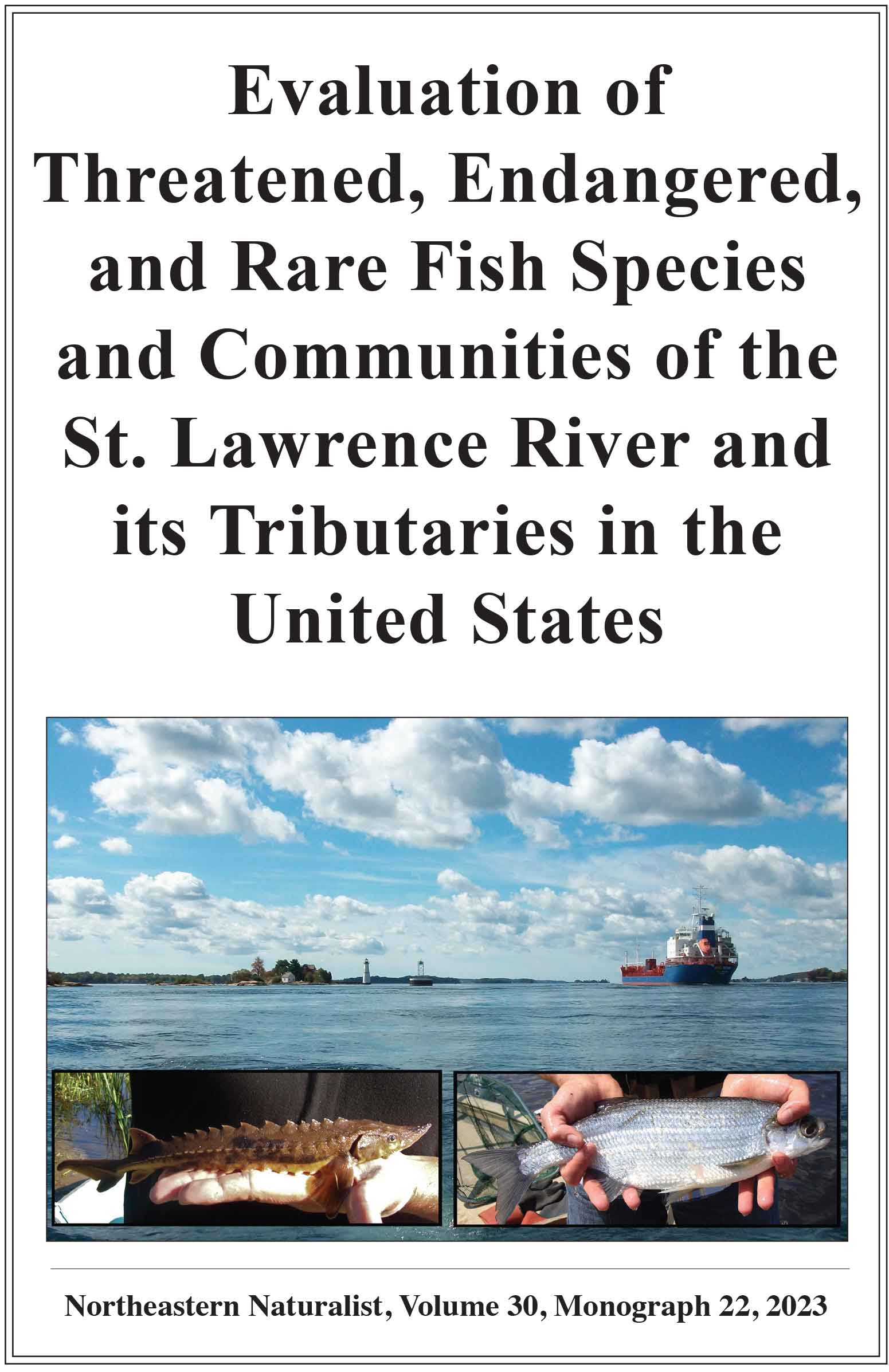
Check out NENA's latest Monograph:
Monograph 22


2016 Northeastern Naturalist Notes Vol. 23, No. 3
N18
M.J. Ravesi, S.J. Tetzlaff, M.C. Allender, and B.A. Kingsbury
Detection of Snake Fungal Disease from a Lampropeltis
triangulum (Eastern Milksnake) in Northern Michigan
Michael J. Ravesi1,*, Sasha J. Tetzlaff
1, Matthew C. Allender2,3, and Bruce A. Kingsbury1
Abstract - An alarming number of cases of Snake Fungal Disease (SFD), a condition frequently
resulting in morbidity and mortality in snakes, have been documented in numerous species across
much of the eastern US. We sampled a skin lesion on the face of a free-ranging Lampropeltis triangulum
(Eastern Milksnake) from the northern Lower Peninsula of Michigan. The lesion tested
positive for Ophidiomyces ophiodiicola, the causative agent of SFD. Our results document the
second species from Michigan known to be infected with SFD. This case adds to the growing body
of literature detailing the distribution of snake species affected, and further indicates that this
pathogen is widespread in the eastern US. We stress the continued need for increased, systematic
sampling efforts to determine the species affected by SFD and the potentially deleterious impacts it
has on snake populations.
Several fungal pathogens have been documented to affect free-ranging wildlife in eastern
North America. Pseudogymnoascus destructans (Blehert & Gargas) Minnis & D.L.
Lindner, the causative agent of White-nose syndrome (WNS; Lorch et al. 2011), has led to
mortality in millions of bats. On a regional scale, for example, WNS caused drastic population
declines in New York (Frick et al. 2010). Similarly, Batrachochytrium dendrobatidis
Longcore, Pessier & D.K. Nichols, which causes Chytridiomycosis, is associated with mass
die-offs and extinction in amphibians globally (Skerratt et al. 2007), including population
declines and extirpations in North America (e.g., Vredenburg et al. 2010). Ophidiomyces
ophiodiicola Guarro, Deanna A. Sutton, Wickes & Rajeev, a fungal pathogen affecting freeranging
North American snakes, is yet another example of an emergent host-specific threat.
Ophidiomyces ophiodiicola has received considerable attention and research in recent
years (reviewed in Allender et al. 2015a). It is related to other species in the Chrysosporium
anamorph Nannizziopsis vriessi (CANV) complex—a group of fungi that are known to
cause dermatitis in numerous reptilian taxa (Sigler et al. 2013). Skin conditions associated
with CANV were initially reported in captive snakes (Nichols et al. 1999, Vissiennon et al.
1999), though similar symptoms were subsequently reported in 2006 from wild Crotalus
horridus (L.) (Timber Rattlesnake) in an imperiled New Hampshire population (Clark et
al. 2011). Researchers also noticed skin lesions and facial swelling in Sistrurus catenatus
(Rafinesque) (Massasauga Rattlesnake) from a population in Carlyle Lake, IL in 2008,
which prompted laboratory analysis confirming the presence of O. ophiodiicola in symptomatic
individuals (Allender et al. 2011). In subsequent years, more cases of Snake Fungal
Disease (SFD) have been reported in various species in geographically disparate locations
throughout eastern North America. Numerous viperids have been documented either with
clinical signs of SFD (Cheatwood et al. 2003, Clark et al. 2011) or confirmed O. ophiodiicola
infection (Allender et al. 2011, 2015a; Smith et al. 2013; Tetzlaff et al. 2015). Simlarly,
1Department of Biology and Environmental Resources Center, Indiana-Purdue University Fort Wayne,
2101 East Coliseum Boulevard, Fort Wayne, IN 46805. 2Wildlife Epidemiology Lab, Department of
Comparative Biosciences, College of Veterinary Medicine, University of Illinois Urbana-Champaign,
2001 South Lincoln Avenue, Urbana, IL 61802. 3Illinois Natural History Survey, Prairie Research
Institute, University of Illinois Urbana-Champaign, 1816 South Oak Street, Champaign, IL 61820.
*Corresponding author - ravemj01@ipfw.edu.
Manuscript Editor: Joseph Milanovich
Notes of the Northeastern Naturalist, Issue 23/3, 2016
N19
2016 Northeastern Naturalist Notes Vol. 23, No. 3
M.J. Ravesi, S.J. Tetzlaff, M.C. Allender, and B.A. Kingsbury
symptoms of SFD or confirmed O. ophiodiicola have also been documented in several
colubrid species (Table 1). Although not the only fungus detected, O. ophiodiicola has been
consistently observed in SFD infections, and it was recently confirmed as the causative
agent of the disease (Allender et al. 2015b, Lorch et al. 2015).
Ophidiomyces causes a range of symptoms including cutaneous ulcers, nodules, hyperkeratosis,
and scale deformities; and it can damage deeper tissue in muscle and bone
(Allender et al. 2011, Lorch et al. 2015, Sigler et al. 2013, Sleeman 2013). Although
O. ophiodiicola infections are nearly always fatal in Massasauga Rattlesnakes, (Allender
et al. 2011, Tetzlaff et al. 2015), Timber Rattlesnakes have been documented to recover
naturally from the disease (Smith et al. 2013).
SFD was first documented in Michigan in 2013 in 2 male Massasauga Rattlesnakes in
the northern Lower Peninsula of Michigan (Tetzlaff et al. 2015). In 2014, SFD persisted
in snakes at that site and was found at 2 additional Massasauga Rattlesnake research
sites in southern Michigan (Allender et al. 2016). Prior to 2015, this species was the only
one in Michigan confirmed to have SFD.
We found a Lampropeltis triangulum (Lacépède) (Eastern Milksnake; snout-to-vent
length = 57.8 cm, tail length = 10.0 cm) on 25 May 2015 in Grayling, MI at 44°40'12.72"N,
84°36'29.46"W, approximately 22 km east of the previously documented cases of SFD in
northern Michigan (Tetzlaff et al. 2015). We captured the individual at a private property
in sandy habitat dominated by Pinus banksiana (Lamb.) (Jack Pine). The snake had obvious
localized thickening of the skin and facial swelling from a discolored, crusty lesion on the
right side of the face consistent with clinical signs of SFD (Allender et al. 2011, Tetzlaff et
al. 2015). We immediately sampled the lesion using sterile, wooden-handled, micro-tipped
Table 1. List of colubrid species in the eastern US either presenting SFD symptoms or confirmed with
O. ophiodiicola infection, from published reports.
Captive
Species State(s) or wild
Pantherophis guttatus (L.) (Red Cornsnake) New York (Sigler et al. 2013) Captive
Nerodia s. sipedon (L.) (Northern Watersnake) Ohio (Sleeman 2013), Wild
Virginia (Guthrie et al. 2016)
Thamnophis sirtalis (L.) (Garter Snake) Florida (Cheatwood et al. 2003) Wild
Thamnophis s. sauritus (L.) (Common Ribbonsnake) Florida (Cheatwood et al. 2003) Wild
Lampropeltis triangulum (Lacépède) (Eastern Milksnake) Wisconsin (Sigler et al. 2013), Wild
New York (Sleeman 2013)
Nerodia clarkia (Baird and Girard) (Saltmarsh Snake) Florida (Sigler et al. 2013) Wild
Coluber constrictor (L.) (Black Racer) Virginia (Guthrie et al. 2016), Wild
Florida (Sleeman 2013)
Pantheropus obsoletus (Holbrook) (Rat Snake) Georgia (Rajeev et al. 2009), Wild
New Jersey (Sleeman 2013)
Thamnophis radix (Baird and Girard) (Plains Garter Illinois (Dolinski et al. 2014) Wild
Snake)
Farancia abacura (Holbrook) (Mud Snake) Georgia (Fenton et al. 2015) Wild
Nerodia fasciata confluens (Blanchard) (Broad-banded Louisiana (Glorioso et al. 2016) Wild
Water Snake)
Farancia erytrograma (Latreille) (Rainbow Snake) Virginia (Guthrie et al. 2016) Wild
Nerodia taxispilota (Holbrook) (Brown Watersnake) Virginia (Guthrie et al. 2016) Wild
2016 Northeastern Naturalist Notes Vol. 23, No. 3
N20
M.J. Ravesi, S.J. Tetzlaff, M.C. Allender, and B.A. Kingsbury
cotton swabs. We collected 2 swab samples from the lesion, which was the only portion of
the snake’s body that we swabbed; the individual was subsequently returned to its original
capture location.
We transferred the samples to 2-ml Eppendorf tubes and stored them at -20 oC until
analysis. We conducted DNA extraction and quantitative PCR amplification (qPCR) tests
for the presence of O. ophiodiicola according to Allender et al. (2015c). Briefly, qPCR was
performed in triplicate on an ABI 7500 real-time thermocycler targeting a 68-bp segment of
the ITS 1 region between the 18S and 5.8S rRNA gene of O. ophiodiicola. The samples were
considered positive because all 3 replicates had a lower cycle-threshold value (Ct = 31.277
± 0.217 SE; 79 fungal copies per reaction) than the lowest detected standard dilution, thus
confirming the presence of O. ophiodiicola.
Confirmed O. ophiodiicola infection (Sigler et al. 2013) or clinical signs of SFD (Sleeman
2013) have previously been observed in Eastern Milksnakes in other portions of its
range. Individual observations such as the one described here continue to be reported for
several species from geographically disparate and noncontiguous regions, providing additional
evidence that this pathogen is widespread in eastern North America and afflicts a
wide breadth of snake species. This observation further emphasizes the need for increased
awareness of SFD symptoms by biologists, veterinarians, and resource managers. If coupled
with a systematic sampling regime (Allender et al. 2013, 2015c), monitoring efforts will aid
in determining the influence of the disease on individual behavior and survivorship, as well
as population-level impacts.
Acknowledgments. This work was conducted under an approved scientific collector’s permit from
the Michigan Department of Natural Resources.
Literature Cited
Allender, M.C., M. Dreslik, S. Wylie, C. Phillips, D.B. Wylie, C. Maddox, M.A. Delaney, and M.J.
Kinsel. 2011. Chrysosporium sp. infection in Eastern Massasauga Rattlesnakes. Emerging Infectious
Diseases 17:2383–2385.
Allender, M.C., M.J. Dreslik, D.B. Wylie, S.J. Wylie, J.W. Scott, C.A. Phillips. 2013. Ongoing health
assessment and prevalence of Chrysosporium in the Eastern Massasauga (Sistrurus catenatus
catenatus). Copeia. 2013:97–102.
Allender, M.C., E. Hileman, J. Moore, and S. Tetzlaff. 2016. Detection of Ophidiomyces, the causative
agent of snake fungal disease, in the Eastern Massasauga (Sistrurus catenatus) in Michigan,
USA. Journal of Wildlife Diseases 52:694–698.
Allender, M.C., D.B. Raudabaugh, F.H. Gleason, and A.N. Miller. 2015a. The natural history, ecology,
and epidemiology of Ophidiomyces ophiodiicola and its potential impact on free-ranging snake
populations. Fungal Ecology 17:187–196.
Allender, M.C., S. Baker, D. Wylie, D. Loper, M.J. Dreslik, C.A. Phillips, C. Maddox, and E.A.
Driskell. 2015b. Development of snake fungal disease after experimental challenge with Ophidiomyces
ophiodiicola in Cottonmouths (Agkistrodon piscivorous). PloS ONE 10:e0140193.
Allender, M.C., D. Bunick, E. Dzhaman, L. Burrus, and C. Maddox 2015c. Development and use of
a real-time polymerase chain-reaction assay for the detection of Ophidiomyces ophiodiicola in
snakes. Journal of Veterinary Diagnostic Investigation 27:217–220.
Cheatwood, J.L., E.R. Jacobson, P.G. May, T.M. Farrell, B.L. Homer, D.A. Samuelson, and J.W.
Kimbrough. 2003. An outbreak of fungal dermatitis and stomatitis in a free-ranging population
of Pigmy Rattlesnakes (Sistrurus miliarius barbouri) in Florida. Journal of Wildlife Diseases
39:329–337.
Clark, R.W., M.N. Marchand, B.J. Clifford, R. Stechert, and S. Stephens. 2011. Decline of an isolated
Timber Rattlesnake (Crotalus horridus) population: Interactions between climate change, disease,
and loss of genetic diversity. Biological Conservation 144:886–891.
N21
2016 Northeastern Naturalist Notes Vol. 23, No. 3
M.J. Ravesi, S.J. Tetzlaff, M.C. Allender, and B.A. Kingsbury
Dolinski, A.C., M.C. Allender, V. Hsiao, and C.W. Maddox. 2014. Systemic Ophidiomyces ophiodiicola
infection in a free-ranging Plains Garter Snake (Thamnophis radix). Journal of Herpetological
Medicine and Surgery 24:7–10.
Fenton, H., L. Last, and J. Goyner-McGuire. 2015. Snake fungal disease in Georgia. Southeastern
Cooperative Wildlife Disease Study, University of Geoergia. SCWDS Briefs 30:4–5.
Frick, W.F., J.F. Pollock, A.C. Hicks, K.E. Langwig, D.S. Reynolds, G.G. Turner, C.M. Butchkoski,
and T.H. Kunz. 2010. An emerging disease causes regional population collapse of a common
North American bat species. Science 329:679–682.
Glorioso, B.M., J.H. Waddle, D.E. Green, and J.M. Lorch. 2016. First documented case of snake
fungal disease in a free-ranging wild snake in Louisiana. South eastern Naturalist: 15:N4–N6.
Guthrie, A.L., S. Knowles, A.E. Ballmann, and J.M. Lorch. 2016. Detection of snake fungal disease
due to Ophidiomyces ophidiodiicola in Virginia, USA. Journal of Wildlife Diseases: 52:143–149.
Lorch, J.M., C.U. Meteyer, M.J. Behr, J.G. Boyles, P.M. Cryan, A.C. Hicks, A.E. Ballmann, J.T.
Coleman, D.N. Redell, and D.M. Reeder. 2011. Experimental infection of bats with Geomyces
destructans causes white-nose syndrome. Nature 480:376–378.
Lorch, J.M., J. Lankton, K. Werner, E.A. Falendysz, K. McCurley, and D.S. Blehert. 2015. Experimental
infection of snakes with Ophidiomyces ophiodiicola causes pathological changes that
typify snake fungal disease. mBio 6:e01534–15.
Nichols, D.K., R.S. Weyant, E.W. Lamirande, L. Sigler, and R.T. Mason. 1999. Fatal mycotic dermatitis
in captive Brown Tree Snakes (Boiga irregularis). Journal of Zoo and Wildlife Medicine
30(1):111–118.
Rajeev, S., D.A. Sutton, B.L. Wickes, D.L. Miller, D. Giri, M. Van Meter, E.H. Thompson, M.G. Rinaldi,
A.M. Romanelli, and J.F. Cano. 2009. Isolation and characterization of a new fungal species,
Chrysosporium ophiodiicola, from a mycotic granuloma of a Black Rat Snake (Elaphe obsoleta
obsoleta). Journal of Clinical Microbiology 47:1264–1268.
Sigler, L., S. Hambleton, and J.A. Paré. 2013. Molecular characterization of reptile pathogens currently
known as members of the Chrysosporium anamorph of Nannizziopsis vriesii complex and relationship
with some human-associated isolates. Journal of Clinical Microbiology 51:3338–3357.
Skerratt, L.F., L. Berger, R. Speare, S. Cashins, K.R. McDonald, A.D. Phillott, H.B. Hines, and N.
Kenyon. 2007. Spread of chytridiomycosis has caused the rapid global decline and extinction of
frogs. EcoHealth 4:125–134.
Sleeman, J. 2013. Snake fungal disease in the United States. Report to Natural Resource/Conservation
Managers. Fungal Disease in the United States. US Geological Survey, Reston, VA.
Smith, C.E., J.E. Edwards, and L.M. Lorch. 2013. Crotalus horridus (Timber Rattlesnake): Fungal
pathogens. Herpetological Review 44:519–520.
Tetzlaff, S.J., M. Allender, M. Ravesi, J. Smith, and B. Kingsbury. 2015. First report of snake fungal
disease from Michigan, USA, involving Massasaugas, Sistrurus catenatus (Rafinesque 1818).
Herpetology Notes 8:31–33.
Vissiennon, T.H., O.F. Schüppel, E. Ullrich, E. Kuijpers, and F.A. Angela. 1999. A disseminated
infection due to Chrysosporium queenslandicum in a Garter Snake (Thamnophis). Mycoses
42:107–110.
Vredenburg, V.T., R.A. Knapp, T.S. Tunstall, and C.J. Briggs. 2010. Dynamics of an emerging disease
drive large-scale amphibian population extinctions. Proceedings of the National Academy of Science
USA 107:9689–9694.