2013 SOUTHEASTERN NATURALIST 12(1):209–216
Heritable Melanism and Parasitic Infection Both Result in
Black-Spotted Mosquitofish
Lisa Horth1,*, David Gauthier1, and Wolfgang Vogelbein2
Abstract - Male Gambusia holbrooki (Eastern Mosquitofish) express a heritable pigmentation
polymorphism: ≈99% of males are silver, and only ≈1% have a melanic, black-spotted
pattern. Sex-linkage, an autosomal modifier, and temperature control the expression of this
heritable melanism. In many teleosts, melanin also accumulates around the site of parasitic
invasion. We have identified black-spot disease in wild mosquitofish from their native habitat.
Here, we demonstrate convergence upon the black-pigmented phenotype through two
means: 1) heritable melanism, and 2) melanic spotting on the silver genotype that results
from infection with immature encysted trematodes. Females are silver and express greater
avoidance of melanic males during mating attempts. The resemblance of the black-spotted
pattern of the melanic genotype to that of silver genotype infected with trematodes may affect
the fitness of melanic males if females perceive them as diseased. Alternatively, females
may shun parasitized silver fish because they resemble the melanic genotype, which is larger
and has a larger mating organ.
Introduction
Mosquitofish are live-bearing fish in the family Poeciliidae (Paige and Burr
1991). Gambusia holbrooki Girard (Eastern Mosquitofish) are native to the
southeastern United States and share a hybrid zone with G. affinis (S.F. Baird &
Girard) (Western Mosquitofish) around Mobile Bay, AL that extends eastward
(Scribner and Avise 1993, Wooten et al. 1988). The wild-type coloration of both
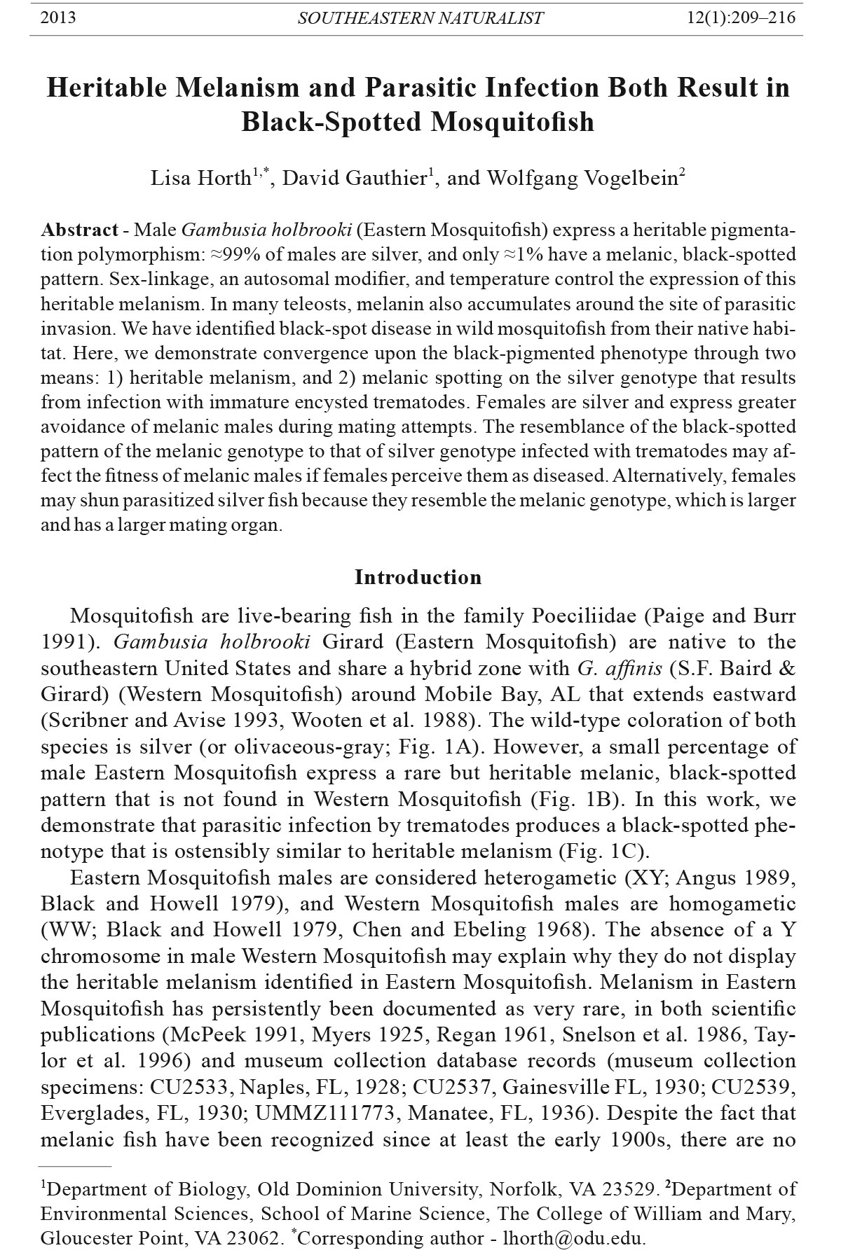
species is silver (or olivaceous-gray; Fig. 1A). However, a small percentage of
male Eastern Mosquitofish express a rare but heritable melanic, black-spotted
pattern that is not found in Western Mosquitofish (Fig. 1B). In this work, we
demonstrate that parasitic infection by trematodes produces a black-spotted phenotype
that is ostensibly similar to heritable melanism (Fig. 1 C).
Eastern Mosquitofish males are considered heterogametic (XY; Angus 1989,
Black and Howell 1979), and Western Mosquitofish males are homogametic
(WW; Black and Howell 1979, Chen and Ebeling 1968). The absence of a Y
chromosome in male Western Mosquitofish may explain why they do not display
the heritable melanism identified in Eastern Mosquitofish. Melanism in Eastern
Mosquitofish has persistently been documented as very rare, in both scientific
publications (McPeek 1991, Myers 1925, Regan 1961, Snelson et al. 1986, Taylor
et al. 1996) and museum collection database records (museum collection
specimens: CU2533, Naples, FL, 1928; CU2537, Gainesville FL, 1930; CU2539,
Everglades, FL, 1930; UMMZ111773, Manatee, FL, 1936). Despite the fact that
melanic fish have been recognized since at least the early 1900s, there are no
1Department of Biology, Old Dominion University, Norfolk, VA 23529. 2Department of
Environmental Sciences, School of Marine Science, The College of William and Mary,
Gloucester Point, VA 23062. *Corresponding author - lhorth@odu.edu.
210 Southeastern Naturalist Vol. 12, No. 1
published accounts addressing the convergence upon the black-spotted phenotype
that results from heritable melanism and parasitic infecti on by trematodes.
“Black spot” is a prevalent freshwater fish disease (Berra and Au 1978) that may
increase in frequency when habitat is degraded (Steedman 1991). Mosquitofish,
and many other fish species, develop it as a result of infection by any of several
digenetic trematodes (or parasitic flatworms with a complex life cycle; Baker
and Bulow 1985; Hoffman 1956, 1967). The name literally refers to the fact that
infected fish appear sprinkled with pin-head-sized black spots on the skin where
parasites are encysted. Black-spot disease is common in many freshwater habitats
of the United States and is thought to retard growth and increase mortality in some
species such as Esox Lucius L. (Northern Pike; Harrison and Hadley 1982) and has
been shown recently to affect behaviors such as shoaling and individual-level association
preferences in Western Mosquitofish (Tobler and Schlupp 2008).
Figure 1. A. The wildtype silver (or olivaceous-gray) male morph of Gambusia holbrooki
(Eastern Mosquitofish). B. The melanic spotted male genotype. C. The silver male parasitized
by trematodes, demonstrating resemblance to the melanic genotype.
2013 L. Horth, D. Gauthier, and W. Vogelbein 211
Here, we report on the convergence upon the black-spotted phenotype in
Eastern Mosquitofish that is produced by two different mechanisms: 1) the production
of heritable melanic pigmentation, and 2) infection of silver-bodied fish
by parasitic trematodes.
Materials and Methods
To evaluate the inheritance of melanism, virgin F1 juvenile fish were reared in
isolation in a 31 °C laboratory after being born to females from one of three wild
populations in Florida (details in Horth 2006). Individual, maturing virgin F1 females
were mated to melanic males from the same population. Additionally, two
populations were reciprocally crossed. One hundred twenty-five matings yielded
over 1000 F2 individuals that were counted to deduce the inheritance pattern of
melanism. F2 fish that did not turn melanic after maturation were moved to an
18 °C cold room to determine whether the decreased temperature would induce
melanic expression. To visually assess melanic deposition in unparasitized fish,
ten silver and ten melanic fish were anaesthetized in MS222, and the dermis of
these fish was observed under a compound microscope at 100x magn ification.
Black-spotted parasitized fish are relatively rare in nature and thus were collected
periodically when identified during routine sampling from four populations
in Florida (Wakulla Springs and Picnic Pond in Wakulla County, Wacissa
River in Jefferson County, and Miami in Dade County) between 1996 and 1999.
Approximately 20 fish were collected and maintained in the lab with the expectation
of performing behavioral observations on them. It was qualitatively noted
that shortly after the invasive plant Hydrilla verticillata (L.f.) Royle (Hydrilla)
was mowed in both Wakulla Springs and the Wacissa River, the local mosquitofish
population size decreased, fish often had small lacerations, and a greater
number of fish appeared diseased.
In 2007, six fish were collected from Miami and Wakulla (3 individuals per population)
and dissected for histological evaluation of pigmentation resulting from
parasitic invasion. Whole fish were preserved in 10% neutral buffered formalin,
decalcified, and processed for paraffin histology according to standard techniques
(Prophet et al. 1994). Thin cross-sections were prepared on a rotary microtome,
stained with hematoxylin and eosin, and examined on an Olympus BX70 compound
microscope. Digital images were generated with an Olympus DP70 camera.
Results and Discussion
Inheritance data demonstrated that Y-linkage and an autosomal modifier largely
explained the heritable melanic black-spotted pigmentation pattern. Melanic penetrance,
or expression, was temperature-sensitive for some populations, and the
life-stage at which melanism was expressed varied by population (details in Horth
2006). For example, in fish from Miami, heritable melanic expression was constitutive
and initiated just a few days after birth in some fish, and after a few weeks
for all fish that expressed melanic pigmentation. In contrast, in fish from Picnic
Pond, melanic expression was induced only after ≈12 weeks of exposure to cold
(18 °C), which also meant post-maturation. In all cases, once deposited, melanic
212 Southeastern Naturalist Vol. 12, No. 1
pigmentation was permanent. Many Picnic Pond fish had only a few spots of small
size. Parasitized fish tended to look quite similar, and often had several (e.g., 5–10
spots) but not more than 20 spots evident externally, as sites of infection.
Compound microscopic examination of the healthy silver genotype at 100x
magnification revealed small dots of melanin distributed in a patterned fashion
across the dermis of the fish. The same basic pattern was also apparent in melanic
fish along with fewer, relatively large blotches of melanin distributed in an
uneven fashion (not pictured, images available from author upon request). The
small dots of pigmentation on both morphs were barely visible by eye, in contrast
with the large blotches of melanin on the melanic genotype.
Upon visual inspection, the phenotype of parasitized silver fish often appeared
nearly indistinguishable from the melanic genotype. One difference, however,
was that the response to the parasite sometimes appeared three dimensional and
resulted in slight protrusion on the surface of the fish at some, but not all, sites of
invasion. This protrusion did not occur for heritable melanic pigmentatio n.
Microscopy also demonstrated that both black-spotted phenotypes resulted
from the deposition of the inert black pigment melanin into the dermis of the
fish. In some parasitized fish, metacercariae (encysted immature parasites) were
present in the hypodermis and even underlying the muscle. The parasite-induced
phenotype resulted from hyperpigmentation surrounding the granulomatous host
cellular response to encysted metacercaria (Noga 2000). This effect was consistent
with the fact that trematode metacercariae often elicit melanin deposition
in finfishes (Roberts 2001, Roberts and Janovy 2005), which typically results in
spots of visible coloration where encysted parasites lie close to the skin surface.
Despite small differences, heritable melanic spots often look the same as
parasite-induced spots, especially on fish from temperature-sensitive populations.
Heritable melanism occasionally also appears as splotches of black
coloration that nearly completely cover a fish. For comparison, when Picnic
Pond fish expressed melanic pigmentation in the 18 °C laboratory, the pigmentation
was expressed as small dots, largely resembling the parasitized phenotype.
In contrast, when Miami fish expressed melanin in the 31 °C laboratory, this
blotchy pigmentation sometimes occurred over a larger area of the body than was
typically covered with parasitism, though there were also fish from Miami that
produced only one or a few melanic spots throughout their lifet ime.
Histopathological evaluation of parasitized fish demonstrated that the grossly
apparent black spots were caused by encysted Neascus metacercariae (Fig. 2). The
host response to the parasite was granulomatous in nature, and highly flattened
epithelioid cells were seen in the inner layer of the cyst (Fig. 3). In some, but not
all parasite-associated granulomas, a layer of chondrocytes existed between the
epithelioid layer and a more distal fibrotic layer of the fish. Hyperpigmentation
Figure 3 (opposite page, bottom). A. Neascus metacercarial cyst in the subcutaneous musculature
of the Eastern Mosquitofish. Metacercaria (Mc) is present within the cyst, and
melanin hyperpigmentation (arrows) is present surrounding the cyst. The epidermis is
indicated by (Epi). B. The cyst wall is made up of epithelioid macrophages (arrowheads),
chondrocytes (*) and a fibrotic layer. Melanin hyperpigmentation is indicated by arrows.
Both scale bars are 50 mm.
2013 L. Horth, D. Gauthier, and W. Vogelbein 213
Figure 2. Neascus metacercarial cyst (Mc) in the hypodermis of the Eastern Mosquitofish.
Flattened epithelioid cells (arrowheads) line the cyst, and hyperpigmentation (arrows)
surrounds the cyst. Scales and scale pockets indicated by (Sc). Scale bar is 50 mm.
214 Southeastern Naturalist Vol. 12, No. 1
due to melanin was present external to the fibrotic layer, and extended between
adjacent myofibers.
A number of strigeoid trematodes cause hyperpigmented spots in the skin
of fishes, including Uvulifer ambloplitis (Hughes) (Black Spot Flatworm). This
diplostomatid trematode has been reported to infect Western Mosquitofish (Spellman
and Johnson 1987). The immature, metacercarial stage of this and related
trematodes is called a neascus metacercaria. It is appropriate to refer to these
parasites as belonging to genus Neascus until the adult parasite may be properly
identified (Roberts and Janovy 2005). Neascus species have a typical digenetic
life cycle where motile miracidia penetrate snails and then produce sporocysts.
The sporocysts produce mobile cercariae that are released by the snails, penetrate
the dermis of fish, and invade the musculature (or other fish tissue). The cercariae
then encyst to the metacercarial stage in the fish tissue. When piscivorous birds
consume infected fish, they ingest the parasite, which then matures and reproduces
in the bird’s intestine. The parasite’s eggs are then extruded into the water
where they hatch into ciliated miracidia.
The genetically based and parasitically induced black-spotted phenotypes
both result from the deposition of insoluble melanin in the skin layers of the
fish. Thus, it is possible that the presence of a parasitic skin infection in silver
males that changes their appearance to black-spotted, could affect the rate at
which females approach melanic genotypes because these males are perceived as
harboring parasites and could potentially be less fit. For example, Eastern Mosquitofish
females were shown to avoid melanic-male mating attempts more than
silver-male mating attempts (Horth 2003).
Alternatively, it females may avoid melanic-male mating attempts for reasons
unrelated to their phenotypic similarity to parasitized fish. If that was the case,
then females may avoid parasitized fish because they resemble melanic genotypes.
In fact, melanic males are larger than silver males and additionally have
relatively larger mating organs (Horth et al. 2010).
We initiated efforts to study female behavior toward parasitized silver, versus
healthy silver and melanic genotypes from all four populations, but parasitized
fish often died in the laboratory not long after collection, indicative of low fitness.
Given that the melanic mosquitofish genotype has been persistently rare in nature,
further investigation is warranted to determine whether females avoid melanic
males because they are considered compromised as high-quality mates due to their
perceived parasite load. If this were the case, the phenomenon might be generalizable
and include other poeciliid species, like Poecilia latipinna (Lesueur) (Sailfin
Mollies) and possibly multiple Xiphophorus (swordtail) species (Axelrod and
Wishnath 1991) which are also comprised of polymorphic populations with melanic
spotted individuals that can be rare. Even unrelated species, like Fundulus
chrysotus (Günther) (Golden Topminnow), that harbor rare melanic individuals
may experience changes in mate choice since they also host trematode parasites
and are sympatric with Eastern Mosquitofish and Sailfin Mollies, where snails and
wading birds are prevalent and where habitat degradation has occurred.
Given a choice between parasitized and non-parasitized individuals, Western
Mosquitofish preferred not to associate with parasitized conspecifics (Tobler
2013 L. Horth, D. Gauthier, and W. Vogelbein 215
and Schlupp 2008). It is noteworthy that one of us (L. Horth) has repeatedly
observed parasitized silver males in nature, but not parasitized melanic fish, in
over 15 years of collection, despite the fact that on most collection trips, melanic
males tended to be dip-netted, measured, and visually scrutinized for assessment
of maturation at which time parasitism could have been detected. Whether the
rarity of melanic fish with parasites is simply a phenomenon of sampling error
(melanic males are very rare), or whether melanin perhaps ironically provides an
anti-parasitic property similar to its purported antibacterial properties in other
organisms such as birds and insects (Burtt and Ichida 2004, Lambrechts et al.
2004) also remains to be determined.
Overall, the convergence upon the black-spotted phenotype of the melanic
genotype and parasitized silver males is a striking natural occurrence. Whether
there are fitness consequences for melanic genotypes resembling parasitized fish,
or parasitized fish resembling melanic genotypes, also remains to be elucidated.
Whether communication regarding parasites is just visual or may also involve
olfaction merits additional research since olfactory cues often contribute to conspecific
knowledge about parasites (Rivière et al. 2009). Additional work on the
effects of parasites and pigmentation polymorphisms may contribute to understanding
why some rare genotypes remain rare, and/or how parasitism may alter
mating behavior dynamics and female choice.
Acknowledgments
Thanks to NSF for funding (DEB 1051015 to L. Horth), to R. Bray for assistance
with photographs at 100x of the dermis of both fish color morphs, and to two anonymous
reviewers who contributed constructive comments.
Literature Cited
Angus, R. 1989. Inheritance of the melanistic pigmentation in the Eastern Mosquitofish.
Journal of Heredity 80:387–392.
Axelrod, H.R., and L. Wischnath. 1991. Swordtails and Platies. TFH Publications, Neptune
City, NJ.
Baker, S.C., and F.J. Bulow. 1985. Effects of black-spot disease on the condition of
Stonerollers, Campostoma anomalum. American Midland Naturalist 114:198–199.
Berra, T.M., and R.-J. Au. 1978. Incidence of black spot disease in fishes in Cedar Fork
Creek, Ohio. Ohio Journal of Science 78:318–22.
Black, D.A., and W.M. Howell. 1979. The North American mosquitofish, Gambusia affinis:
A unique case in sex-chromosome evolution. Copeia 1979:509–513.
Burtt, E.H., and J.M. Ichida. 2004. Gloger’s rule, feather-degrading bacteria, and color
variation among Song Sparrows. The Condor 106:681–686.
Chen, T.R., and A.W. Ebeling. 1968. Karyological evidence of female heterogamety in
the Mosquitofish, Gambusia affinis. Copeia 1968:70–75.
Harrison, E.J., and W.F. Hadley. 1982. Possible effects of black-spot disease on Northern
Pike. Transactions of the American Fisheries Society 111:106–109.
Hoffman, G.L. 1956. The life cycle of Crassiphiala bulboglossa (Trematoda: Strigeida).
Development of the metacercaria and cyst, and effect on the fish hosts. Journal of
Parasitology 42:435–444.
Hoffman, G.L. 1967. Parasites of North American Freshwater Fishes. University of California
Press, Berkeley, CA. 486 pp.
216 Southeastern Naturalist Vol. 12, No. 1
Horth, L. 2003. Melanic body-color and aggressive mating behavior are correlated traits
in male Mosquitofish (Gambusia holbrooki). Proceedings of the Royal Society of
London 270:1033–1040.
Horth, L. 2006. A sex-linked allele, autosomal modifiers, and temperature dependence
appear to regulate melanism in male Mosquitofish (Gambusia holbrooki). Journal of
Experimental Biology 209:4938–4945.
Horth, L., C. Binckley, R. Wilk, P. Reddy and A. Reddy. 2010. Color, body size, and
genitalia size are correlated traits in Eastern Mosquitofish (Gambusia holbrooki).
Copeia 2010(2):196–202.
Lambrechts, L., J.M. Vulule, and J.C. Koella. 2004. Genetic correlation between melanization
and antibacterial immune responses in a natural population of the malaria
vector Anopheles gambiae. Evolution 58:2377–2381.
McPeek, M.A. 1991. Mechanisms of sexual selection operating on body size in the Mosquitofish
(Gambusia holbrooki). Behavioral Ecology 3:1–12.
Myers, G.S. 1925. Concerning melanomorphism (spp.) in killifishes. Copeia 137:105–107.
Noga, E.J. 2000. Fish Disease: Diagnosis and Treatment. Iowa State University Press,
Ames, IA.
Paige, L.M., and B.M. Burr. 1991. A Field Guide to Freshwater Fishes. Houghton Miflin
Company. New York, NY. 417 pp.
Prophet, E.B., B. Mills, J.B. Arrington, and L.H. Sobin. 1994. Laboratory Methods in
Histotechnology. Armed Forces Institute of Pathology, 7th Edition. McGraw Hill. New
York, NY.
Regan, J.D. 1961. Melanism in the poecillid fish Gambusia affinis (Baird & Girard).
American Midland Naturalist 65:139–143.
Rivière, S., L. Challet, D. Fluegge, M. Spehr, and I. Rodriguez. 2009. Formyl peptide
receptor-like proteins are a novel family of vomeronasal chemosensors. Nature
468:574–577.
Roberts, L.S. 2001. Fish Pathology. W.B. Saunders. London, UK.
Roberts, L.S., and J. Janovy, Jr. 2005. Foundations of Parasitology. McGraw Hill, New
York, NY.
Scribner, K.T., and J.C. Avise. 1993. Cytonuclear genetic architecture in mosquitofish
populations and the possible roles of introgressive hybridization. Molecular Ecology
2:139–149.
Snelson, F.F., R.E. Smith, and M.R. Bolt. 1986. A melanistic female mosquitofish.
American Midland Naturalist 115:413–415.
Spellman, S.J., and A.D. Johnson. 1987. In vitro excystment of the Black Spot Trematode,
Uvulifer ambloplitis (Trematoda: Diplostomatidae). International Journal for
Parasitology 17:897–902.
Steedman, R.J. 1991. Occurrence and environmental correlates of black spot disease in
stream fishes near Toronto, Ontario. Transactions of the American Fisheries Society
120:494–499.
Taylor, S.A., E. Burt, G. Hammond, and K. Releya. 1996. Female mosquitofish prefer normally
pigmented males to melanistic males. Comparative Psychology 110:260–266.
Tobler, M. and I. Schlupp. 2008. Influence of black spot disease on shoaling behaviour
in female Western Mosquitofish, Gambusia affinis (Poeciliidae, Teleostei). Environmental
Biology of Fishes 81:29–34.
Wooten, M.C., K.T. Scribner, and M.H. Smith. 1988. Genetic variability and systematics
of Gambusia in the southeastern United States. Copeia 1988:283–289.


 The Southeastern Naturalist is a peer-reviewed journal that covers all aspects of natural history within the southeastern United States. We welcome research articles, summary review papers, and observational notes.