2016 Southeastern Naturalist Notes Vol. 15, No. 1
N4
B.M. Glorioso, J.H. Waddle, D.E. Green, and J.M. Lorch
First Documented Case of Snake Fungal Disease in a Freeranging
Wild Snake in Louisiana
Brad M. Glorioso1,*, J. Hardin Waddle1, D. Earl Green2, and Jeffrey M. Lorch2
Abstract - Snake fungal disease (SFD) is a recently documented mycotic disease characterized by
scabs or crusty scales, subcutaneous nodules, abnormal molting, cloudiness of the eyes (not associated
with molting), and localized thickening or crusting of the skin. SFD has been documented in many
species in the Eastern and Midwestern United States within the last decade. SFD has proven lethal in
many snakes, and the disease is recognized as an emerging threat to wild snake populations. Herein
we describe the first documented case of SFD in Louisiana in a f ree-ranging wild snake.
Recently, fungal pathogens have been implicated in severe population declines in vertebrates
such as bats (Blehert et al. 2009) and anurans (Skerratt et al. 2007). Wild snakes
have also been affected by fungal pathogens, notably Ophidiomyces ophiodiicola (Guarro,
Deanna A. Sutton, Wickes, & Rajeev) Sigler, Hambleton, & Paré, which is believed to
be the causative agent of snake fungal disease (SFD; Allender et al. 2011, 2015; Smith et
al. 2013).
Cases of SFD in Sistrurus catenatus (Rafinesque) (Massasauga) are reported to be 100%
lethal (Allender et al. 2011, Tetzlaff et al. 2015), whereas 2 infected Crotalus horridus L.
(Timber Rattlesnake) had improved health over 10 weeks in captivity without treatment to
the point of one being asymptomatic (Smith et al. 2013). Little is known about the prevalence,
severity, and population-level impacts of SFD (Smith et al. 2013).
Recently, as awareness of SFD has increased, the Amphibian Research and Monitoring
Initiative team at the Wetland and Aquatic Research Center (WARC) has been
opportunistically examining all captured snakes for clinical signs of the disease during
the course of amphibian monitoring at locations in Louisiana and Texas. One location
where we routinely see snakes with clinical signs of infection is Cypress Island Preserve.
The property, better known as Lake Martin, is owned by The Nature Conservancy, and
is located in St. Martin Parish, LA, about 8 km south of Breaux Bridge. On 26 March
2015, we observed a juvenile Nerodia fasciata confluens (Blanchard) (Broad-banded
Watersnake) coiled and basking alongside the trail on the southwestern part of the lake at
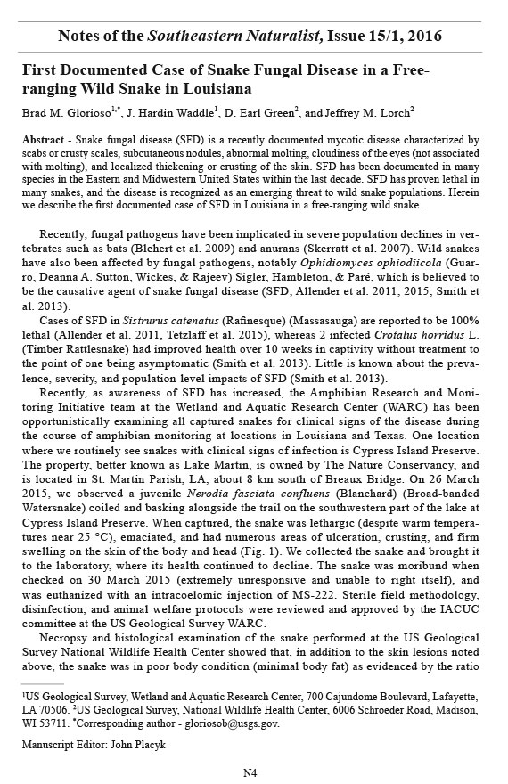
Cypress Island Preserve. When captured, the snake was lethargic (despite warm temperatures
near 25 °C), emaciated, and had numerous areas of ulceration, crusting, and firm
swelling on the skin of the body and head (Fig. 1). We collected the snake and brought it
to the laboratory, where its health continued to decline. The snake was moribund when
checked on 30 March 2015 (extremely unresponsive and unable to right itself), and
was euthanized with an intracoelomic injection of MS-222. Sterile field methodology,
disinfection, and animal welfare protocols were reviewed and approved by the IACUC
committee at the US Geological Survey WARC.
Necropsy and histological examination of the snake performed at the US Geological
Survey National Wildlife Health Center showed that, in addition to the skin lesions noted
above, the snake was in poor body condition (minimal body fat) as evidenced by the ratio
1US Geological Survey, Wetland and Aquatic Research Center, 700 Cajundome Boulevard, Lafayette,
LA 70506. 2US Geological Survey, National Wildlife Health Center, 6006 Schroeder Road, Madison,
WI 53711. *Corresponding author - gloriosob@usgs.gov.
Manuscript Editor: John Placyk
Notes of the Southeastern Naturalist, Issue 15/1, 2016
N5
2016 Southeastern Naturalist Notes Vol. 15, No. 1
B.M. Glorioso, J.H. Waddle, D.E. Green, and J.M. Lorch
Figure 1. A juvenile Broad-banded Watersnake (Nerodia fasciata confluens) collected during a survey
at Cypress Island Preserve, St. Martin Parish, LA, which tested positive for Ophidiomyces ophiodiicola.
The snake exhibited ulceration of the skin on the head (A), several crusty ventral scales (B), and
numerous nodules overlaid by areas of roughened skin on the dorsal surface (C, D).
of its snout–vent length (266 mm) to its weight (10.2 g). On microscopic examination,
fungi consistent with O. ophiodiicola were observed in the skin lesions, and O. ophiodiicola
was isolated in culture from multiple skin lesions. Fungal identification was confirmed by
sequencing the entire internal transcribed spacer region of the ribosomal RNA gene as described
in Bohuski et al. (2015).
This is the first documented occurrence of SFD in a free-ranging wild snake in
Louisiana, and one of the few documented cases in the United States of SFD in juvenile
snakes. Clinical signs consistent with the disease have been observed in snakes from
many areas of Louisiana in the last few years, and we do not believe the snake in this
report represents an isolated case. In some areas where we capture snakes frequently, we
have not found individuals with clinical signs of the disease, although this does not preclude
its presence. We plan on conducting additional surveillance and diagnostic testing
for SFD in wild snakes in Louisiana to help elucidate the epidemiology and ecological
significance of this emerging disease.
Acknowledgments. We thank Anne E. Ballmann and the necropsy and diagnostic laboratory
technicians at the US Geological Survey NWHC for assistance in coordinating
sample submission and processing samples. We appreciate the field assistance received
from Lindy Muse, Sidney Godfrey, and Brome McCreary. Matt Pardue and Kacy King of
the TNC provided permitting and logistical assistance. Animal husbandry methods were
reviewed and authorized by the Institutional Animal Care and Use Committee (IACUC)
2016 Southeastern Naturalist Notes Vol. 15, No. 1
N6
B.M. Glorioso, J.H. Waddle, D.E. Green, and J.M. Lorch
at the US Geological Survey Wetland and Aquatic Research Center. Any use of trade,
firm, or product names is for descriptive purposes only and does not imply endorsement
by the US Government.
Literature Cited
Allender, M.C., M. Dreslik, S. Wylie, C. Phillips, D.B. Wylie, C. Maddox, M.A. Delaney, and M.J.
Kinsel. 2011. Chrysosporium sp. infection in eastern massasauga rattlesnakes. Emerging Infectious
Diseases 17:2383–2384.
Allender, M.C., D.B. Raudabaugh, F.H. Gleason, and A.N. Miller. 2015. The natural history, ecology,
and epidemiology of Ophidiomyces ophiodiicola and its potential impact on free-ranging snake
populations. Fungal Ecology 17:187–196.
Blehert, D.S., A.C. Hicks, M.J. Behr, C.U. Meteyer, B.M. Berlowski-Zier, E.L. Buckles, J.T.H. Coleman,
S.R. Darling, A. Gargas, R. Niver, J.C. Okoniewski, R.J. Rudd, and W.B. Stone. 2009. Bat
white-nose syndrome: An emerging fungal pathogen? Science 323:227.
Bohuski, E., J.M. Lorch, K.M. Griffin, and D.S. Blehert. 2015. TaqMan real-time polymerase chain
reaction for detection of Ophidiomyces ophiodiicola, the fungus associated with snake fungal
disease. BMC Veterinary Research 11:95.
Skerratt, L.F., L. Berger, R. Speare, S. Cashins, K.R. McDonald, A.D. Phillott, H.B. Hines, and N.
Kenyon. 2007. Spread of chytridiomycosis has caused the rapid global decline and extinction of
frogs. EcoHealth 4:125–134.
Smith, C.E., J. Edwards, and J.M. Lorch. 2013. Crotalus horridus (Timber Rattlesnake): Fungal
pathogens. Herpetological Review 44:519–520.
Tetzlaff, S., M. Allender, M. Ravesi, J. Smith, and B. Kingsbury. 2015. First report of snake fungal
disease from Michigan, USA involving Massasaugas, Sistrurus catenatus (Rafinesque 1818).
Herpetology Notes 8:31–33.


 The Southeastern Naturalist is a peer-reviewed journal that covers all aspects of natural history within the southeastern United States. We welcome research articles, summary review papers, and observational notes.