2008 NORTHEASTERN NATURALIST 15(2):303–308
Pelage Spotting and Staining in Eastern Moles
(Scalopus aquaticus)
Ava A. Kamm1, George A. Feldhamer1,*, and John D. Reeve1
Abstract - We quantified relative extent of pelage staining in Scalopus aquaticus
(eastern mole) as an indicator of scent-gland marking, and evaluated whether staining
was associated with colored pelage spots and patches often prevalent on the snout
and ventral surface of individuals. Moles were collected from southern Illinois (n =
91) and from Cincinnati, OH (n = 152). Adult moles scent-marked more than juveniles,
but pelage staining was independent of breeding season for males and females.
Pelage spotting occurred in 33.7% of the sample and was not associated with pelage
staining from glandular secretions, as has been suggested by some previous investigators.
Pelage spots were most prevalent on the ventral surface. Ventral spotting
occurred more often in males than females (P < 0.001). Mean area of ventral spots
was 2.81 cm2 with no differences in area related to sex or age.
Introduction
Like many other talpids, Scalopus aquaticus Linnaeus (eastern moles)
rarely are studied and are difficult to observe directly because they live underground.
Adults generally are solitary except during the breeding season.
Home ranges may overlap, although more than one animal rarely is found in
a tunnel system (Harvey 1976). Olfactory communication is more effective
for moles than visual or auditory signaling (Gorman and Stone 1990). Moles
use urine and anal secretions at distinct sites throughout their area where
such marking is most likely to be encountered to alert conspecifics that an
area is occupied. The abdominal fur of moles often is stained brown from
glandular secretions. In addition to direct marking with urine, preputial secretions
also are deposited in tunnels of Talpa europaea Linnaeus (European
moles) as they move (Gorman and Stone 1990).
Pelage of the eastern mole is short and velvety; it is gray in the northern
part of the range and brown or tan in the southern and western part (Whitaker
1996). Orange, yellow, or olive tints on the chin and other parts of the pelage
occur in eastern moles and many other North American species (Cockrum and
Meinkoth 1942, Hartman and Yates 2003, Miller 1921). Colored fur often occurs
where sudoriferous glands are abundant such as the dorsal snout, chin,
breast, abdomen, wrists, and the perineal region. Eadie (1954) reported that
the orange spots were a product of the sudoriferous and perineal glands, and
that spotting was more pronounced in males during the breeding season. Conversely,
Jackson (1915) and Carraway and Verts (1991) believed that pelage
spotting in moles was a genetic factor. Our objectives were to determine if age,
gender, breeding season, body mass, pregnancy, or location affect the presence
1Department of Zoology, Southern Illinois University, Carbondale, IL 62901-6501.
*Corresponding author - feldhamer@zoology.siu.edu.
304 Northeastern Naturalist Vol. 15, No. 2
and intensity of pelage staining and spotting in eastern moles, and to determine
if pelage staining affects the presence and intensity of pelage spotting.
Methods and Materials
Data collection
Eastern moles were collected from southern Illinois (approximately
37.72°N, 89.20°W) and the Cincinnati, OH (39.16°N, 84.46°W) area from
October 2004 until May 2005. All specimens were collected with kill traps
(Victor® mole traps) primarily by professional mole trappers at both locales.
All specimens were stored at about 10 °C until postmortem examinations were
conducted. Specimens were weighed to the nearest gram on a Hanson Dietetic
Scale Model 144. Total length, tail length, and hind-foot length were recorded,
and skulls were removed and cleaned using dermestid beetle larvae. The reproductive
tracts of females and the testes of males were removed and weighed to
the nearest 0.01 g on a Denver Instrument Digital Scale to help assess the reproductive
status of individuals. In addition, females were categorized either
as pregnant or not pregnant by visual inspection of the uterus and ovaries.
Sex, date and location of collection, and age were recorded for each specimen,
although sex could not be determined for all specimens. Yearly age classes
from juvenile through 5 years were determined based on wear criteria of the
upper cheek teeth (Hartman 1995) of cleaned skulls. We classified juveniles as
“young” (those collected April through June and that likely were within their
natal area), or “old” (collected July through December and dispersed from
their natal area) based on reproductive data, tooth-wear characteristics of the
last upper molar, and date of capture.
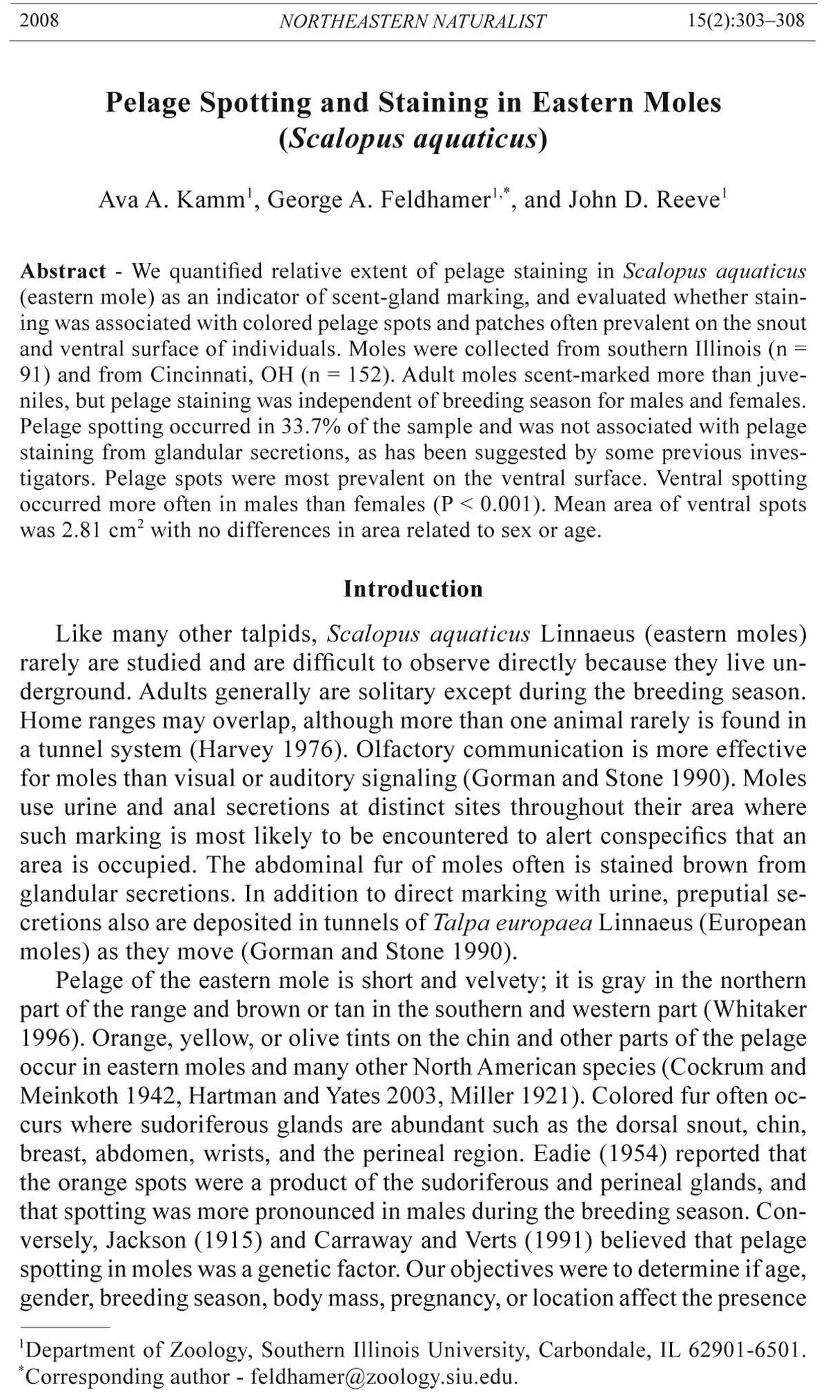
We assessed two aspects of pelage color variation in eastern moles. The
brown wash that often appears on the mid-ventral line, snout, and chin of
animals caused by glandular secretions we termed “pelage staining.” The
distinct patches of fur primarily on the venter (Fig. 1) and snout that range
in color from white to bright orange we considered “pelage spotting.” Pelage
spotting and staining were quantified using a Munsell Color Chart based
on the intensity of color. To quantify staining, the hue, value, and chroma
(Munsell Color 1975) were combined into one of three categories: strong
(= 2), light (= 1), or none (= 0). A pelage stain value from 0 to 2 was assigned
to each area on the body where staining occurred—snout, chin, chest, and
genitals. Ventral staining results in a different color than the flanks and the
dorsum of the animal. If the midline staining was ≥3 Munsell values different
than the flanks and dorsum, the staining was considered “strong.” If the
staining was 2 chroma values different than the flanks and dorsum, the staining
was considered “light.” If the midline was the same color as the flanks
and the dorsum, the animal was considered to have no staining. Pelage staining
values from the snout, chin, chest, and genital areas were combined to
form a score between 0 and 8.
Pelage spotting could also occur on the snout, chin, or chest, occasionally
extending to the genitals of an individual. Pelage spotting was categorized as
“strong” if the Munsell value was yellow-orange to brown-orange and was
2008 A.A. Kamm, G.A. Feldhamer, and J.D. Reeve 305
coded as 2 for analyses, “light” if the value appeared yellow or beige (code
= 1), and “no color” if the fur appeared white (code = 0). Individuals with
no spotting were coded as -1.
Statistical analyses
SAS 9.1 for Windows (SAS Institute, Inc., Cary, NC) was used for
statistical analyses. General linear models were used to test the effect of independent
variables (sex, season, body mass, age, location, and pregnancy)
on the dependent variable total pelage staining. A general linear model was
also used to test the effect of the independent variables sex, season, body
mass, age, location, and pelage staining on the presence or absence and color
intensity of pelage spotting. Season was considered as pre-breeding (October,
November, and December), breeding (January, February, March, April,
and May) and post-breeding (June, July, August, and September). Age was
recorded as juvenile (individuals less than 1 year of age) or adult (individuals ≥1
year of age). Location either was Illinois or Ohio.
General linear models were constructed with males and females combined
and the variable “pregnancy” excluded. After conducting this initial
test, it was determined that sex had no significant effect on pelage staining,
but did have a significant effect on pelage spotting. Thus, the general linear
models for pelage staining were conducted again, separating males and females
to include the independent variable pregnancy in the females’ model.
To reduce the number of variables in the general linear models, those that
had P > 0.25 were eliminated. Variables with P ≤ 0.25 then were tested for
significant interactions. Interactions with P > 0.05 were not included in the
final model. The adjusted Least Squares Means were calculated for each
significant variable in the model, and the Tukey-Kramer method was used
to test for significant differences between the adjusted means for each group
found significant. The upper and lower 95% confidence intervals for each
Figure 1. Large orange pelage spot on the venter of an eastern mole.
306 Northeastern Naturalist Vol. 15, No. 2
adjusted mean also were calculated. Data were not transformed because
residuals were normally distributed.
Results
A total of 243 moles was collected. We collected 91 individuals from
southern Illinois (19 juvenile males, 34 adult males, 13 juvenile females, and
25 adult females) and 152 moles from Cincinnati, OH (24 juvenile males, 39
adult males, 25 juvenile females, and 64 adult females). Mean mass of testes
peaked in February (mean = 1.16 g ± 0.10), consistent with anticipated peak
breeding based on latitude (Gorman and Stone 1990). We found most adult
females were pregnant in March.
Pelage stains
For males, age had a significant effect on pelage staining (P < 0.0001),
with adults having more staining than juveniles (Table 1). The adjusted mean
value for staining in adults was 6.7 ± 0.9, whereas that for juveniles was
2.8 ± 1.3. The general linear model explained 61% of the variation in male
pelage staining. Adult females had more pelage staining (P < 0.0003) than
did juvenile females (Table 1); the adjusted mean staining was 4.9 ± 0.7 for
adults and 2.5 ± 1.3 for juveniles. In contrast to our findings for males, the
general linear model for females explained only 17% of the variation. The
month of capture was known for 70 of our 81 juveniles. “Young” juveniles
(n = 50) had a significantly lower mean staining value (mean = 0.70 ± 0.33)
than “old” juveniles (n = 20; mean = 3.85 ± 0.53; t = 5.04, P less than 0.0001).
Pelage spots
Of 243 moles, 82 (33.7%) had pelage spots in one or more areas. Spotting
occurred on the snout and chin, and rarely on the wrist or top of the head. Spotting
usually was orange and was most pronounced on the chest and abdomen. A
single contiguous chest/abdominal spot occurred in 45 individuals, whereas 8
others had two to four separate ventral spots. The mean area of ventral spotting
was 2.81 cm2 (SD = 4.65) and ranged from 0.04 to 25.0 cm2. There was no difference
in the mean area of ventral spots between males (3.22 cm2) and females
(1.59 cm2) (F = 1.31, d.f. = 49, P = 0.26). Likewise, there was no relationship
between the area of ventral spots and age (F = 0.354, R2 = 0.009). Given the
Table 1. Variables affecting pelage staining in Scalopus aquaticus (eastern mole) collected in
southern Illinois and Cincinnati, OH from 2004 to 2005.
Source of variation d.f. F-value P-value
Males
Season 2 1.67 0.20
Body mass 1 3.66 0.06
Age 1 21.92 less than 0.0001
Location 1 2.66 0.11
Age x season 2 0.90 0.41
Age x location 1 3.21 0.08
Females
Age 1 13.69 0.0003
Pregnancy 1 3.65 0.06
2008 A.A. Kamm, G.A. Feldhamer, and J.D. Reeve 307
observed sex ratio of our sample for specimens where sex was determined
(1M:1.11F), we found significantly more males (n = 36) with ventral pelage
spots than females (n = 15; χ2 = 11.75, d.f. = 49, P < 0.001). None of the variables
in the general linear model for pelage spotting was significant (Table 2),
and the model explained only 0.05% of the variation in the data.
Discussion
Pelage stains
The primary factor that affected pelage staining in males and females was
age; adults had significantly more staining than juveniles. Juveniles, especially
those that were captured during the first 6 months of the year, likely
would not have left their natal territories yet and would not need to scentmark
to establish their own territories. Eadie (1954) found that juveniles
had no anal gland staining until 4 months after birth. This is consistent with
differences we found in juveniles depending on whether they were collected
earlier or later in the year. Nonetheless, the adult female model had very
little explanatory power. Thus, factors investigated in this study did little to
explain what variables affect scent-marking in females.
Surprisingly, staining was not significantly affected by season. This suggests
that the frequency that eastern moles scent-mark is the same regardless
of the season. If there is an increase in scent-marking during the breeding season,
we did not detect it based on pelage staining. Although the composition of
glandular secretions differs during the breeding season, at least in European
moles (Khazanehdari et al. 1996), the amount of scent-marking by eastern
moles throughout the year may consistently display territorial information.
Similar to Eadie (1954), we found that adults had more pelage staining
than juveniles. Conversely, he found that males had more staining than females,
and that adults had more staining in the breeding season than at other
times of the year. His findings may have been biased by his methods. He
graded the areas of staining as “strong,” “light,” “trace,” or “none” for each
specimen and designated arbitrary values on a numbered scale of 1–10 for
those 4 grades. Furthermore, Eadie did not use a color chart to measure the
levels of staining, nor did he include staining of the genital area in his study;
he also considered orange pelage spots as staining.
Pelage spots
The general linear model results for pelage spotting only explained 0.05%
of the variation in the data. Therefore, the model had no biological significance
Table 2. General linear model for effects of pelage stains on pelage spots in Scalopus aquaticus
(eastern moles) collected 2004–2005. Both sexes were combined.
Source of variation d.f. F-value P-value
Staining 1 0.17 0.68
Sex 1 3.15 0.08
Season 2 1.30 0.27
Body mass 1 0.21 0.65
Age 1 0.20 0.66
308 Northeastern Naturalist Vol. 15, No. 2
in terms of factors investigated in our study. Carraway and Verts (1991) also
found an absence of seasonality in the color intensity or presence of pelage
spots in Scapanus townsendii Bachman (Townsend’s mole). As would be expected
if spotting was a genetic trait, we found age or glandular staining had no
effect on spotting. We conclude, as did Carraway and Verts (1991), that pelage
spotting was not due to glandular secretions but is genetically controlled.
Hartman and Yates (2003) noted that yellow, orange, or olive pelage spotting
is a widespread phenomenon in several genera of North American moles
in addition to Scalopus, including Condylura, Parascalops, and Scapanus.
It is difficult to ascribe any adaptive significance to brightly colored ventral
pelage in a subterranean species such as the eastern mole. Nonetheless, such
coloration must not reduce personal fitness either, given its common occurrence
within and among populations of eastern moles and other talpids.
Acknowledgments
A previous draft of the manuscript benefited from the input of L.N. Carraway
and an anonymous reviewer. We thank local mole trappers for supplying most of the
specimens for this research. We especially thank Greg Hartman for his insight on age
determination of moles and editorial assistance on the manuscript. Karen Jones, Department
of Animal Science, provided helpful insight throughout this study. Support
was provided through the Department of Zoology, Southern Illinois University.
Literature Cited
Carraway, L.N., and B.J. Verts. 1991. Pattern and color aberrations in pelages of
Scapanus townsendii. Northwest Science 65:16–21.
Cockrum, E.L., and N.A. Meinkoth. 1942. Abnormal coloration in the prairie mole.
Journal of Mammalogy 23:451.
Eadie, W.R. 1954. Skin gland activity and pelage descriptions in moles. Journal of
Mammalogy 35:186–196.
Gorman, M.L., and R.D. Stone. 1990. The Natural History of Moles. Cornell University
Press, Ithaca, NY. 138 pp.
Hartman, G.D. 1995. Age determination, age structure, and longevity in the mole,
Scalopus aquaticus (Mammalia: Insectivora). Journal of Zoology, London
237:107–122.
Hartman, G.D., and T.L. Yates. 2003. Moles, Talpidae. Pp. 30–55, In G.A. Feldhamer,
B.C. Thompson, and J.A. Chapman (Eds.). Wild Mammals of North
America: Biology, Management, and Conservation. Johns Hopkins University
Press, Baltimore, MD. 1216 pp.
Harvey, M.J. 1976. Home range, movements, and diel activity of the eastern mole,
Scalopus aquaticus. American Midland Naturalist 95:436–445.
Jackson, H.H.T. 1915. A review of the American moles. North American Fauna
38:1–100.
Khazanehdari, C., A.J. Buglass, and J.S. Waterhouse. 1996. Anal gland secretion of
European mole: Volatile constituents and significance in territorial maintenance.
Journal of Chemical Ecology 22:383–392.
Miller, L. 1921. The coat color of moles. Journal of Mammalogy 2:163–166.
Munsell Color. 1975. Munsell soil color charts. Kollmorgen Corp., Baltimore, MD.
Whitaker, J.O. Jr. 1996. National Audubon Society Field Guide to North American
Mammals. Chanticleer Press Inc., New York, NY. 745 pp.