Detection of a Babesia Species in a Bobcat from Georgia
Barbara C. Shock1,2,*, J. Mitchell Lockhart3, Adam J. Birkenheuer4,
and Michael J. Yabsley1,2
Abstract - We describe the first detection of a Babesia sp. in a Lynx rufus (Bobcat). The Bobcat
was from Georgia and was coinfected with Cytauxzoon felis and a Sarcocystis sp. The Babesia
species was closely related to Babesia sp. “Coco”, a parasite previously only detected in Canis
familiaris (Domestic Dog). The only other Babesia sp. in North America that infects felids is a
novel Babesia species in Puma concolor coryi (Florida Puma). The low prevalence of this Babesia
(less than 1%) in Bobcats suggests that they are not the normal host or reservoir and this may have been
an incidental infection.
Piroplasms (genera Babesia, Theileria, and Cytauxzoon) are tick-transmitted
apicomplexan parasites which infect a wide range of mammals and birds worldwide (Criado-
Fornelio et al. 2004). Species of all three genera infect erythrocytes, but in contrast to
Theileria and Cytauxzoon, Babesia spp. do not have an extra-erythrocytic stage (Criado-
Fornelio et al. 2004). Numerous species of piroplasms are important disease-causing agents
for veterinary species, and Babesia are notable in that many species are zoonotic. Disease
caused by Babesia is rare among wildlife, but disease can develop during stressful periods,
after co-infection with immunosuppressive viruses, or when infections occur in aberrant
hosts (e.g., Panthera leo L. [Lions] from Africa infected with a natural Babesia species but
diseased when exposed to drought and coinfected with Canine Distemper Virus, or when exotic
Rangifer tarandus L. [Reindeer] in the northeastern United States become infected with
Babesia species native to the area; Bartlett et al. 2009, Munson et al. 2008).
Currently, only two piroplasms have been reported from felines in North America,
Cytauxzoon felis Kier in Felis catus L. (Domestic Cat), Lynx rufus Schreber (Bobcat),
and Puma concolor L. (Puma) from the eastern United States, and a novel Babesia species
in Puma concolor coryi Bangs (Florida Puma) from southern Florida (Glenn et al.
1983, Yabsley et al. 2006). During a surveillance study (Shock et al. 2012) on wildlife
reservoirs of C. felis involving Bobcats (n = 799) and Pumas (n = 49) from thirteen
states (Florida, Georgia, Kansas, Kentucky, Louisiana, Missouri, North Carolina, North
Dakota, Ohio, Oklahoma, Pennsylvania, South Carolina, and West Virginia), a Babesia
species was detected in a single female Bobcat from Thomas County, GA (n = 143; 0.7%).
This is the first report of a Babesia sp. infection in a Bobcat and is only the second felidinfecting
Babesia species reported in North America.
The internal transcribed spacer (ITS)-1 region was amplified using a nested PCR that
amplifies all known piroplasms (Bostrom et al. 2008). Briefly, for primary amplification,
5 ml of DNA was added to 20 ml of a master mix containing 10 mM Tris-Cl (pH 8.3), 50
mM KCl, 1.5 mM MgCl2, 0.2 mM each dNTP (Promega, Madison, WI), 2.5 units GoTaq®
Flexi DNA Polymerase (Promega), and 0.8 mM of primers ITS-15C (5’-CGATCGAGTGATCCGGTGAATTA)
and ITS-13B (5’-GCTGCGTCCTTCATCGTTGTG). Cycling
1Southeastern Cooperative Wildlife Disease Study, 586 D.W. Brooks Drive, Wildlife Health Building,
College of Veterinary Medicine, University of Georgia, Athens, GA, 30602. 2Warnell School
of Forestry and Natural Resources, 180 E Green Street, University of Georgia, Athens, GA 30602.
3Biology Department, Valdosta State University, 1500 N Patterson, Department of Biology, Room
2035, Valdosta, GA 31698. 4NC State College of Veterinary Medicine, 4700 Hillsborough Street,
Raleigh, NC 27606. *Corresponding author - barbarashock@gmail.com.
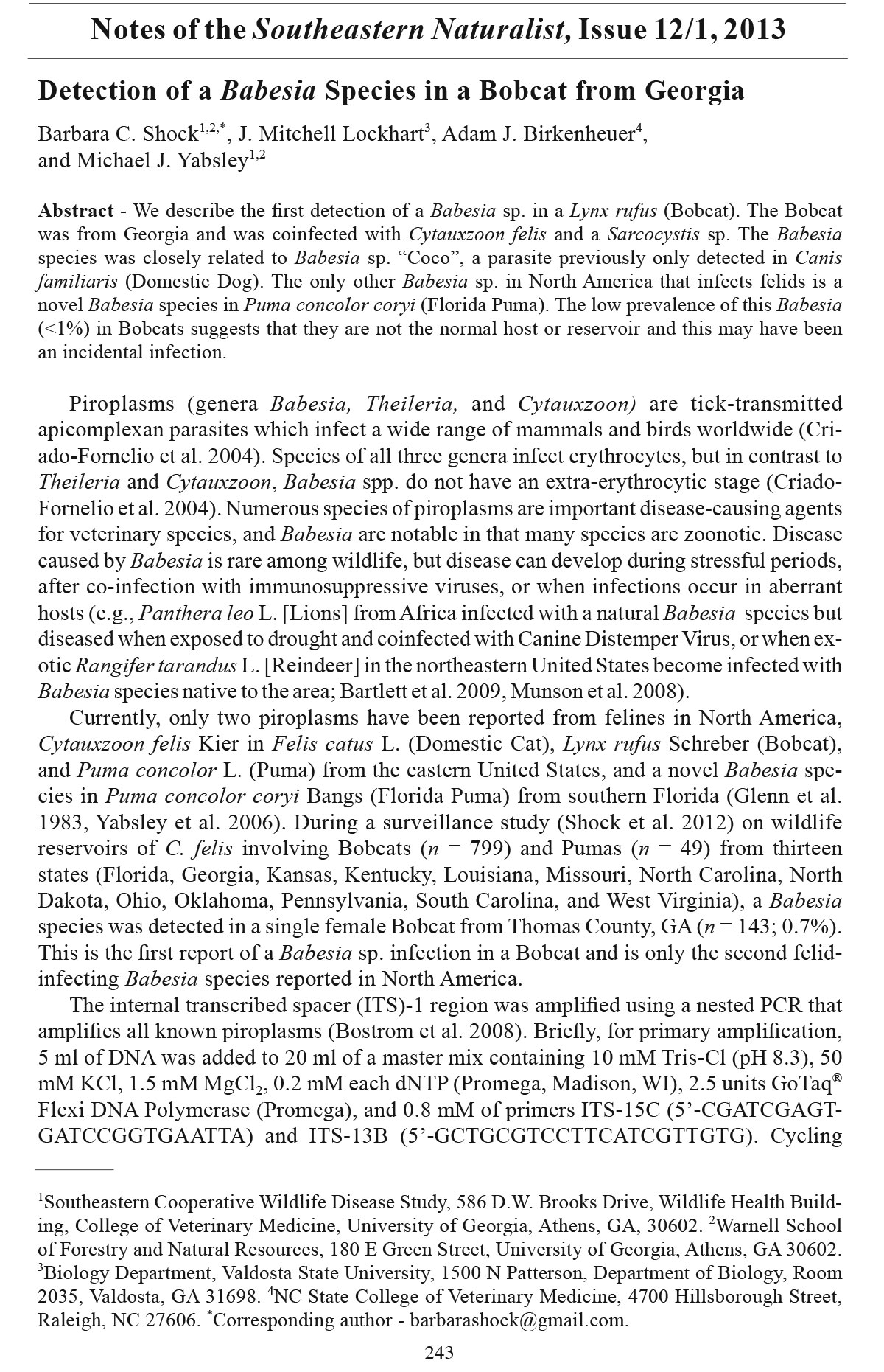
Notes of the Southeastern Naturalist, Issue 12/1, 2013
243
244 Southeastern Naturalist Vol. 12, No. 1
parameters were 94 °C for 1 min followed by 35 cycles of 94 °C for 30 sec, 52 °C for 30
sec, 72 °C for 1 min, and a final extension at 72 °C for 5 min. For the nested PCR, 1 ml
of primary product was used as a template in a 25-ml reaction containing the same PCR
components except inclusion of primers ITS-15D (5’-AAGGAAGGAGAAGTCGTAACAAGG)
and ITS-13C (5’-TTGTGTGAGCCAAGACATCCA). The cycling parameters
were the same as the primary reaction except the annealing temp erature was 49 °C.
To prevent and detect contamination, primary and secondary amplification, and product
analysis were done in separate dedicated areas. A negative water control was included
in each set of DNA extraction, and one water control was included in each set of primary
and secondary PCR reactions. The amplicon from the positive Bobcat was purified with
a Qiagen gel extraction kit (Germantown, MD) and bi-directionally sequenced at the
University of Georgia Integrated Biotechnology Laboratory (Athens, GA).
Sequence analysis of the ITS-1 region (601 bp) indicated that the greatest similarity
(92%) was with a novel large Babesia sp. “Coco’” that was first identified in a Canis
familiaris L. (Domestic Dog) from North Carolina in 2002 (GenBank accession number:
AY618928; Fig. 1; Birkenheuer et al. 2004). The phylogenetic relationship based on
ITS1 between this Babesia and other Babesia spp. is similar to the relationship between
Babesia sp. “Coco” and other Babesia spp. based on analysis of the 18S rRNA gene
(Birkenheuer et al. 2004). The only difference between the Bobcat Babesia sequence
and Babesia sp. “Coco” was the presence of a 45-bp insert in the Bobcat Babesia sp. at
nucleotide site 434. Outside the insert region, bases 1–434 and 435 to 557 of Babesia sp.
“Coco” (EU109720) were 100% similar to our Bobcat Babesia. Thus, we believe that this
Figure 1. Phylogenetic relationships between Babesia spp. inferred from internal transcribed spacer
(ITS)-1 rRNA region sequences.
2013 Southeastern Naturalist Notes 245
Bobcat Babesia sp. represents a variant of Babesia sp. “Coco” and not a novel Babesia
sp., although additional studies are needed to definitively determine the con-specificity of
these two Babesia spp. Insertions and deletions are common in the ITS regions of other
piroplasms (Aktas et al. 2007, Brown et al. 2009, Shock et al. 2012). Attempts to amplify
other gene targets failed as the Bobcat was also co-infected with C. felis, and other targets
(e.g., 18S rRNA gene and ITS-2) were positive but when sequenced were confirmed to be
C .felis. These data highlight the need to utilize multiple gene targets when conducting
pathogen surveillance. Unfortunately, a blood smear was not available from the Babesia
sp.-infected Bobcat so no morphologic data is available.
Babesia sp. “Coco” has only previously been reported from immunosuppressed Domestic
Dogs (Sikorski et al. 2010), so in an effort to better understand why this Bobcat
was infected, we conducted additional pathogen screening using the limited samples
available from this trapper-harvested animal. Serum from the Bobcat was negative for
Feline Immunodeficiency Virus (FIV) antibodies and Feline Leukemia Virus (FeLV) antigens
(IDEXX, Westbrook, ME). A low antibody titer (1:10) for Feline Panleukopenia
Virus was detected (Animal Health Diagnostic Center, Cornell University, Ithaca, NY),
which was not interpreted as an infection. PCR testing for other pathogens revealed that
the Bobcat was positive for “Candidatus Bartonella volans” and was negative for hemoplasmas
(Cadenas et al. 2008, Jensen et al. 2001). Histological examination of available
tissues was unrewarding due to advanced autolysis, but Sarcocystis sp. cysts were observed
in muscle tissue. All of these findings were considered incidental.
Currently, little is known about the natural history of Babesia sp. “Coco” and the
Babesia sp. detected in the Bobcat from Georgia. Babesia sp. “Coco” was first reported
from an immunosuppressed dog undergoing chemotherapy for lymphoma (Birkenheuer
et al., 2004). Since the initial detection, eight additional canine infections have been reported
from dogs, all with a travel history to Mid-Atlantic states. Six of these dogs were
splenectomized, and two were immunosuppressed due to oncolytic drugs (Birkenheuer et
al. 2004, Holman et al. 2009, Sikorski et al. 2010). At least 5 of the 9 dogs infected with
Babesia sp. “Coco” had a history of tick exposure, and at least one sustained bites to the
face, a risk factor for other Babesia sp., such as B. gibsoni Patton (Holman et al. 2009,
Sikorski et al. 2010, Yeagley et al. 2009).
Worldwide, several Babesia spp. have been reported from felids including B. herpailuri
Dennig and B. pantherae Dennig and Brocklesby from wild felids in Africa,
B. felis Davis and B. leo Penzhorn from domestic cats and wild felids in Africa, B. cati
Mudaliar in Domestic Cats from India, B. canis canis Uilenberg from domestic cats in
Spain, B. canis presentii Baneth from Domestic Cats in Israel; a Babesia sp. from Domestic
Cats in Portugal, (Baneth et al. 2004, Criado-Fornelio et al. 2004, Penzhorn et
al. 2004). In the United States, the only previous report of a Babesia species in a felid
is a novel Babesia sp. from Florida Pumas (Yabsley et al. 2006). The Puma Babesia
sp. appears to be restricted to Florida Pumas because, in the current study, infections
were not detected in 49 Pumas from Texas, Louisiana, Georgia, or North Dakota.
Interestingly, the Babesia sp. from the Florida Puma is a small piroplasm morphologically
and is indistinguishable from C. felis, whereas Babesia sp. “Coco” is a large
Babesia. In addition, the two feline-infecting Babesia species from North America are
easily distinguished based on sequence analysis of the ITS-1 region (B.C. Shock et al.,
unpublished data).
In summary, a Babesia sp. closely related to Babesia sp. “Coco” was detected in a
single Bobcat from Georgia, which is the first report of Babesia infection of a Bobcat
and the second report of Babesia in felids from North America. Bobcats likely do not
246 Southeastern Naturalist Vol. 12, No. 1
represent a natural host of this Babesia sp.; thus, additional surveillance studies are
needed to understand the natural host of this parasite.
Acknowledgments. The authors thank numerous personnel from state agencies who
assisted with the collection of felid samples. This study was primarily funded by the Morris
Animal Foundation (DO8FE-003), and additional support was provided by the Federal
Aid to Wildlife Restoration Act (50 Stat. 917) and through sponsorship from fish and
wildlife agencies in Alabama, Arkansas, Florida, Georgia, Kansas, Kentucky, Louisiana,
Maryland, Mississippi, Missouri, North Carolina, Oklahoma, Pennsylvania Puerto Rico,
South Carolina, Tennessee, Virginia, and West Virginia.
Literature Cited
Aktas, M., K.G. Bendele, K. Altay, N. Dumanli, M. Tsuji, and P.J. Holman. 2007. Sequence polymorphism
in the ribosomal DNA internal transcribed spaces differs among Theileria species.
Veterinary Parasitology 147:221–230.
Baneth, G., M.J. Kenny, S. Tasker, Y. Anug, V. Shkap, A. Levy, and S.E. Shaw. 2004. Infection with
a proposed new subspecies of Babesia canis, Babesia canis subsp. presentii, in domestic cats.
Journal of Clinical Microbiology 42:99–105.
Bartlett, S.L., N. Abou-Madi, J.B. Messick, A. Birkenheuer, and G.V. Kollias. 2009. Diagnosis and
treatment of Babesia odocoilei in captive Reindeer (Rangifer tarandus tarandus) and recognition
of three novel host species. Journal of Zoo and Wildlife Medicine 40:152–159.
Birkenheuer, A.J., J. Neel, D. Ruslander, M.G. Levy, and E.B. Breitschwerdt. 2004. Detection and
molecular characterization of a novel large Babesia species in a dog. Veterinary Parasitology
124:151–160.
Bostrom, B., C. Wolf, C. Greene, and D.S. Peterson. 2008. Sequence conservation in the rRNA
first internal transcribed spacer region of Babesia gibsoni genotype Asia isolates. Veterinary
Parasitology 152:152–157.
Brown, H.M., R.D. Berghaus, K.S. Latimer, J.O. Britt, P.M. Rakich, and D.S. Peterson. 2009. Genetic
variability of Cytauxzoon felis from 88 infected domestic cats in Arkansas and Georgia.
Journal of Veterinary Diagnostic Investigation 21:59–63.
Cadenas, M.B., J. Bradley, R.G. Maggi, M. Takara, B.C. Hegarty, and E.B. Breitschwerdt. 2008.
Molecular characterization of Bartonella vinsonii subsp. berkhoffii Genotype III. Journal of
Clinical Microbiology 46:1858–1860.
Criado-Fornelio A., M.A. Gonzalez-del-Rio, A. Buling-Sarana, and J.C. Barba-Carretero. 2004.
The “expanding universe” of piroplasms. Veterinary Parasitology 119:337–345.
Glenn B.L., A.A. Kocan, and E.F. Blouin. 1983. Cytauxzoonosis in Bobcats. Journal of the American
Veterinary Medical Association 183:1155–1158.
Holman, P.J., B.B. Backlund, A.L. Wilcox, R. Stone, A.L. Stricklin, and K.E. Bardin. 2009. Detection
of a large unnamed Babesia piroplasm originally identified in dogs in North Carolina in
a dog with no history of travel to that state. Journal of the American Veterinary Medical Association
235:851–854.
Jensen, W.A., M.R. Lappin, S. Kamkar, W.J. Reagan. 2001. Use of a polymerase chain reaction
assay to detect and differentiate two strains of Haemobartonella felis in naturally infected cats.
American Journal of Veterinary Research 62:604–608.
Munson L, K.A. Terio, R. Kock, T. Mlengeya, M.E. Roelke, E. Dubovi, B. Summers, A.R. Sinclair,
and C. Packer. 2008. Climate extremes promote fatal co-infections during canine distemper
epidemics in African Lions. PLoS One 3(6):e2545.
Penzhorn, B.L., T. Schoeman, and L.S. Jacobson. 2004. Feline babesiosis in South Africa: A review.
Annuls of the New York Academy of Science 1026:183–186.
Shock, B.C., A.J. Birkenheuer, L.L. Patton, C. Olfenbuttel, J. Beringer, D.M. Grove, M. Peek, J.W.
Butfiloski, D.W. Hughes, J.M. Lockhart, M.W. Cunningham, H.M. Brown, D.S. Peterson, and
M.J. Yabsley. 2012. Variation in the ITS-1 and ITS-2 rRNA genomic regions of Cytauxzoon
felis from Bobcats and Pumas in the eastern United States and comparison with sequences from
Domestic Cats. Veterinary Parasitology [Epub ahead of print].
2013 Southeastern Naturalist Notes 247
Sikorski, L.E., A.J. Birkenheuer, M.K. Holowaychuk, A.L. McCleary-Wheeler, J.M. Davis, and
M.P. Littman. 2010. Babesiosis caused by a large Babesia species in 7 immunocompromised
dogs. Journal of Veterinary Internal Medicine 24:127–131.
Yabsley M.J., S.M. Murphy, and M.W. Cunningham. 2006. Molecular detection and characterization
of Cytauxzoon felis and a Babesia species in Cougars from Florida. Journal of Wildlife
Diseases 42:366–374.
Yeagley, T.J., M.V. Reichard, J.E. Hempstead, K.E. Allen, L.M. Parsons, M.A. White, S.E. Little,
and J.H. Meinkoth. 2009. Detection of Babesia gibsoni and the canine small Babesia “Spanish
isolate” in blood samples obtained from dogs confiscated from dogfighting operations. Journal
of the American Veterinary Medical Association 235:535–539.


 The Southeastern Naturalist is a peer-reviewed journal that covers all aspects of natural history within the southeastern United States. We welcome research articles, summary review papers, and observational notes.